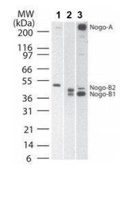
All lanes : Anti-Nogo A+B antibody (ab47085) at 1/2000 dilutionLane 1 : Human brain tissue lysateLane 2 : Mouse brain tissue lysateLane 3 : Rat brain tissue lysateObserved band size : 41,43,48-50,>200 kDa (why is the actual band size different from the predicted?)

ab47085 staining Nogo A+B in African Green Monkey COS-7 cells by ICC/IF (Immunocytochemistry/immunofluorescence). Cells were fixed with paraformaldehyde, permeabilized with 0.5% Triton X-100 and blocked with 3% BSA for 1 hour at 23°C. Samples were incubated with primary antibody (1/500 in PBS-BSA) for 1 hour at 23°C. An Alexa Fluor® 488-conjugated Goat anti-rabbit IgG polyclonal (1/1000) was used as the secondary antibody.See Abreview

Immunocytochemistry stainning for Nogo A+B in Cor1 neural stem cells from mouse using Rabbit polyclonal to Nogo A+B (ab47085; 1/200 incubated for 2h at RT); Immunoreactivity was detected in cell-body as well as in neurites.


