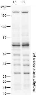
All lanes : Anti-Nogo antibody (ab32298) at 1 µg/mlLane 1 : Rat Hippocampus Tissue Lysate Lane 2 : Mouse Hippocampus Tissue LysateLysates/proteins at 20 µg per lane.SecondaryGoat Anti-Rabbit IgG H&L (HRP) preadsorbed (ab97080) at 1/5000 dilutionPerformed under reducing conditions.

Immunofluorescent staining for Nogo in rat brain cerebellum using Rabbit polyclonal to Nogo (ab32298; 1/300 incubated for 18h at RT); immunoreactivity was detected in most brain areas. The staining is quite diffuse on cell bodies, axons and dendrites. The picture was acquired using X20 objective. Protocol details: Rats were intracardially perfused with 4% paraformaldehyde. Whole brain tissue was post-fixed overnight in the same fixative, and cryoprotected in 20% sucrose and frozen in OCT. 30µm coronal sections were cut by cyrostat for use in fre floating IHC. Secondary antibody Alexa fluor 488 1/1000 was incubated for 2 hours at room temperature.

Immunocytochemistry stainning for Nogo in Cor1 neural stem cells from mouse using Rabbit polyclonal to Nogo (ab32298; 1/200 incubated for 1h at RT); Immunoreactivity was detected in cell-body as well as in neurites.

ab32298 staining Nogo in human SH-SY5Y cells by Immunocytochemistry/ Immunofluorescence.Cells were fixed in paraformaldehyde, blocked with 10% serum for 20 minutes at 24°C, then incubated with ab32298 at a 1/200 dilution for 16 hours at 4°C. The secondary used was an Alexa-Fluor 488 conjugated donkey anti-rabbit polyclonal used at a 1/1000 dilution. Counterstained with Hoechst 33258 (blue).See Abreview


