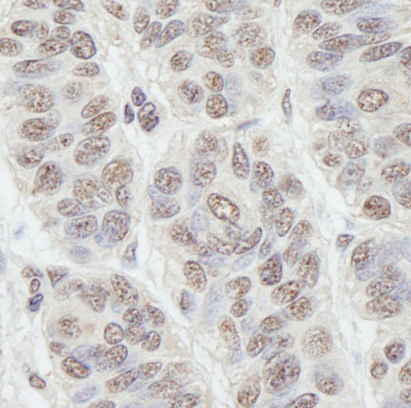
Immunohistochemistry (Formalin/PFA-fixed paraffin-embedded sections) analysis of human skin carcinoma tissue labelling Nucleostemin with ab70346 at 1/1000 (0.2µg/ml). Detection: DAB.

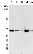
All lanes : Anti-Nucleostemin antibody (ab70346) at 0.04 µg/mlLane 1 : HeLa whole cell lysate at 50 µgLane 2 : HeLa whole cell lysate at 15 µgLane 3 : HeLa whole cell lysate at 5 µgLane 4 : 293T whole cell lysate at 50 µgLane 5 : mouse 3T3 whole cell lysate at 50 µg

Detection of Nucleostemin by Western Blot of Immunprecipitate. All lanes: ab70346 at 1µg/ml staining Nucleostemin in HeLa whole cell lysates immunoprecipitated at 3µg/mg lysate (1 mg/IP; 20% of IP loaded/lane) using the following antibodies: Lane 1: an irrelevant antibody. Lane 2: ab70345 Lane 3: ab70346. Detection: Chemiluminescence with exposure time of 3 seconds

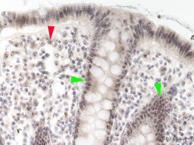
Immunohistochemistical detection of Nucleostemin with ab70346 on formaldehyde-fixed paraffin-embedded human colon sections. Antigen retrieval step: heat mediated in citric acid pH6 buffer. Blocking step: 1% BSA for 10 mins @ rt°C. Primary antibody ab70346 incubated at 1/50 for 16 hours in TBS/BSA/azide. Secondary antibody: anti-rabbit IgG conjugated to biotin (1/200). The intestinal epithelium is the most rapidly self-renewing tissue in adult mammals. Stem cell markers have been observed in cycling columnar cells at the crypt base (Nature 449, 1003-1007; 2007). The cells (nuclei of the simple columnar epithelium of the upper mucosa) immunoreactive for Nucleostemin in this image (green arrowheads) anatomically correspond to the same cells expected to be Lgr5 positivite. A red arrowhead indicates nucleolar positivity in one of many cell nuclei in the Lamina propria of the mucosa (this area is not known for stem cell positSee Abreview

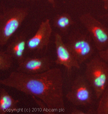
ICC/IF image of ab70346 stained HeLa cells. The cells were 4% formaldehyde fixed (10 min) and then incubated in 1%BSA / 10% normal goat serum / 0.3M glycine in 0.1% PBS-Tween for 1h to permeabilise the cells and block non-specific protein-protein interactions. The cells were then incubated with the antibody (ab70346, 5µg/ml) overnight at +4°C. The secondary antibody (green) was Alexa Fluor® 488 goat anti-rabbit IgG (H+L) used at a 1/1000 dilution for 1h. Alexa Fluor® 594 WGA was used to label plasma membranes (red) at a 1/200 dilution for 1h. DAPI was used to stain the cell nuclei (blue) at a concentration of 1.43µM.


