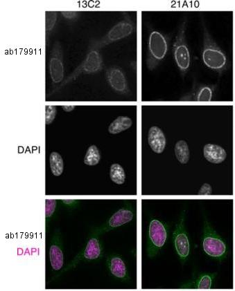
Immunofluorescence analysis of methanol-fixed HeLa cells, labeling NUP98 using two different clones of ab179911 at 0.5 µg/ml, followed by Alexa Fluor 488-conjugated anti-mouse lgG (green) at 4 µg/ml. DAPI was used to stain DNA (magenta). Upper and middle panels correspond to black-and-white images while the bottom panel represents merged colored images.
![Lane 1 : Anti-NUP98 antibody [13C2 + 21A10] - BSA and Azide free (ab179911) at 0.4 µg/ml (13C2 clone)Lane 2 : Anti-NUP98 antibody [13C2 + 21A10] - BSA and Azide free (ab179911) at 0.4 µg/ml (21A10 clone)Lane 1 : HeLa cell extractLane 2 : HeLa cell extractSecondaryHRP-labeled anti-mouse IgG at 0.4 µg/mldeveloped using the ECL technique](http://www.bioprodhub.com/system/product_images/ab_products/2/sub_4/3088_ab179911-209522-ab179911WB11.jpg)
Lane 1 : Anti-NUP98 antibody [13C2 + 21A10] - BSA and Azide free (ab179911) at 0.4 µg/ml (13C2 clone)Lane 2 : Anti-NUP98 antibody [13C2 + 21A10] - BSA and Azide free (ab179911) at 0.4 µg/ml (21A10 clone)Lane 1 : HeLa cell extractLane 2 : HeLa cell extractSecondaryHRP-labeled anti-mouse IgG at 0.4 µg/mldeveloped using the ECL technique

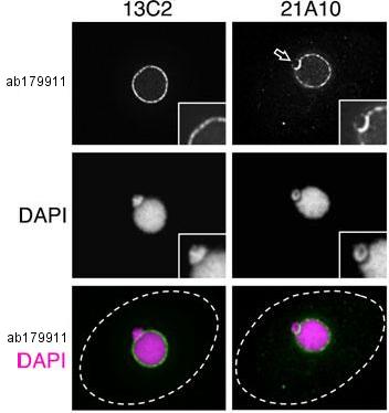
Immunofluorescence analysis of methanol-fixed Tetrahymena thermophila cells, labeling NUP98 using two different clones of ab179911 at 0.5 µg/ml, followed by Alexa Fluor 488-conjugated anti-mouse lgG (green) at 4 µg/ml. DAPI was used to stain DNA (magenta). Upper and middle panels correspond to black-and-white images while the bottom panel represents merged images. Dotted lines represent the outlines of cells. The open arrow indicates the micronucleus. Insets are magnified images showing the position of the micronucleus.
![Lanes 1 & 4 : Anti-NUP98 antibody [13C2 + 21A10] - BSA and Azide free (ab179911) at 2 µg/ml (13C2 clone)Lanes 2 - 3 : Anti-NUP98 antibody [13C2 + 21A10] - BSA and Azide free (ab179911) at 2 µg/ml (21A10 clone)Lane 1 : Tetrahymena thermophila cell extractLane 2 : Tetrahymena thermophila cell extractLane 3 : Tetrahymena thermophila cell extractLane 4 : Cell extract from Tetrahymena thermophila expressing endogenously NUP98 fused to a fluorescence proteindeveloped using the ECL technique](http://www.bioprodhub.com/system/product_images/ab_products/2/sub_4/3090_ab179911-209520-ab179911WB21.jpg)
Lanes 1 & 4 : Anti-NUP98 antibody [13C2 + 21A10] - BSA and Azide free (ab179911) at 2 µg/ml (13C2 clone)Lanes 2 - 3 : Anti-NUP98 antibody [13C2 + 21A10] - BSA and Azide free (ab179911) at 2 µg/ml (21A10 clone)Lane 1 : Tetrahymena thermophila cell extractLane 2 : Tetrahymena thermophila cell extractLane 3 : Tetrahymena thermophila cell extractLane 4 : Cell extract from Tetrahymena thermophila expressing endogenously NUP98 fused to a fluorescence proteindeveloped using the ECL technique

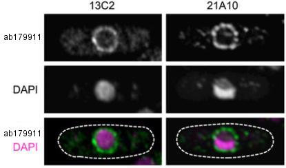
Immunofluorescence analysis of formaldehyde-fixed, zymolyase-treated, S. pombe cells, labeling NUP98 using two different clones of ab179911 at 10 µg/ml, followed by Alexa Fluor 488-conjugated anti-mouse lgG (green). DAPI was used to stain DNA (magenta). Upper and middle panels correspond to black-and-white images while the bottom panel represents merged images. Dotted lines represent the outlines of cells.
![Lanes 1 - 2 : Anti-NUP98 antibody [13C2 + 21A10] - BSA and Azide free (ab179911) at 2 µg/ml (13C2 clone)Lanes 3 - 4 : Anti-NUP98 antibody [13C2 + 21A10] - BSA and Azide free (ab179911) at 2 µg/ml (21A10 clone)Lane 1 : Cell extract from S. pombe wild typeLane 2 : Cell extract from S. pombe expressing endogenously NUP98 fused to a fluorescence proteinLane 3 : Cell extract from S. pombe wild typeLane 4 : Cell extract from S. pombe expressing endogenously NUP98 fused to a fluorescence proteindeveloped using the ECL technique](http://www.bioprodhub.com/system/product_images/ab_products/2/sub_4/3092_ab179911-209518-ab179911WB3.jpg)
Lanes 1 - 2 : Anti-NUP98 antibody [13C2 + 21A10] - BSA and Azide free (ab179911) at 2 µg/ml (13C2 clone)Lanes 3 - 4 : Anti-NUP98 antibody [13C2 + 21A10] - BSA and Azide free (ab179911) at 2 µg/ml (21A10 clone)Lane 1 : Cell extract from S. pombe wild typeLane 2 : Cell extract from S. pombe expressing endogenously NUP98 fused to a fluorescence proteinLane 3 : Cell extract from S. pombe wild typeLane 4 : Cell extract from S. pombe expressing endogenously NUP98 fused to a fluorescence proteindeveloped using the ECL technique

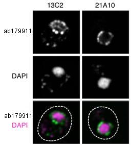
Immunofluorescence analysis of formaldehyde-fixed, zymolyase-treated, S. cerevisiae cells, labeling NUP98 using two different clones of ab179911 at 10 µg/ml, followed by Alexa Fluor 488-conjugated anti-mouse lgG (green). DAPI was used to stain DNA (magenta). Upper and middle panels correspond to black-and-white images while the bottom panel represents merged images. Dotted lines represent the outlines of cells.
![Lane 1 : Anti-NUP98 antibody [13C2 + 21A10] - BSA and Azide free (ab179911) at 2 µg/ml (13C2 clone)Lane 2 : Anti-NUP98 antibody [13C2 + 21A10] - BSA and Azide free (ab179911) at 2 µg/ml (21A10 clone)Lane 1 : S. cerevisiae cell extractLane 2 : S. cerevisiae cell extractdeveloped using the ECL technique](http://www.bioprodhub.com/system/product_images/ab_products/2/sub_4/3094_ab179911-209516-ab179911WB4.jpg)
Lane 1 : Anti-NUP98 antibody [13C2 + 21A10] - BSA and Azide free (ab179911) at 2 µg/ml (13C2 clone)Lane 2 : Anti-NUP98 antibody [13C2 + 21A10] - BSA and Azide free (ab179911) at 2 µg/ml (21A10 clone)Lane 1 : S. cerevisiae cell extractLane 2 : S. cerevisiae cell extractdeveloped using the ECL technique

Summary of the suitability of ab179911 clones for immunological applications. IF: indirect immunofluorescence staining; WB: Western blotting analysis.

![Lane 1 : Anti-NUP98 antibody [13C2 + 21A10] - BSA and Azide free (ab179911) at 0.4 µg/ml (13C2 clone)Lane 2 : Anti-NUP98 antibody [13C2 + 21A10] - BSA and Azide free (ab179911) at 0.4 µg/ml (21A10 clone)Lane 1 : HeLa cell extractLane 2 : HeLa cell extractSecondaryHRP-labeled anti-mouse IgG at 0.4 µg/mldeveloped using the ECL technique](http://www.bioprodhub.com/system/product_images/ab_products/2/sub_4/3088_ab179911-209522-ab179911WB11.jpg)

![Lanes 1 & 4 : Anti-NUP98 antibody [13C2 + 21A10] - BSA and Azide free (ab179911) at 2 µg/ml (13C2 clone)Lanes 2 - 3 : Anti-NUP98 antibody [13C2 + 21A10] - BSA and Azide free (ab179911) at 2 µg/ml (21A10 clone)Lane 1 : Tetrahymena thermophila cell extractLane 2 : Tetrahymena thermophila cell extractLane 3 : Tetrahymena thermophila cell extractLane 4 : Cell extract from Tetrahymena thermophila expressing endogenously NUP98 fused to a fluorescence proteindeveloped using the ECL technique](http://www.bioprodhub.com/system/product_images/ab_products/2/sub_4/3090_ab179911-209520-ab179911WB21.jpg)

![Lanes 1 - 2 : Anti-NUP98 antibody [13C2 + 21A10] - BSA and Azide free (ab179911) at 2 µg/ml (13C2 clone)Lanes 3 - 4 : Anti-NUP98 antibody [13C2 + 21A10] - BSA and Azide free (ab179911) at 2 µg/ml (21A10 clone)Lane 1 : Cell extract from S. pombe wild typeLane 2 : Cell extract from S. pombe expressing endogenously NUP98 fused to a fluorescence proteinLane 3 : Cell extract from S. pombe wild typeLane 4 : Cell extract from S. pombe expressing endogenously NUP98 fused to a fluorescence proteindeveloped using the ECL technique](http://www.bioprodhub.com/system/product_images/ab_products/2/sub_4/3092_ab179911-209518-ab179911WB3.jpg)

![Lane 1 : Anti-NUP98 antibody [13C2 + 21A10] - BSA and Azide free (ab179911) at 2 µg/ml (13C2 clone)Lane 2 : Anti-NUP98 antibody [13C2 + 21A10] - BSA and Azide free (ab179911) at 2 µg/ml (21A10 clone)Lane 1 : S. cerevisiae cell extractLane 2 : S. cerevisiae cell extractdeveloped using the ECL technique](http://www.bioprodhub.com/system/product_images/ab_products/2/sub_4/3094_ab179911-209516-ab179911WB4.jpg)
