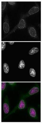
Immunofluorescent analysis of HeLa cells labeling NUP98 with ab179894 at 0.5 µg/ml in PBS (top panel). DAPI staining (middle panel). 4 μg/ml of Alexa488-labeled anti-mouse IgG was used as secondary antibody. The color image represents merged images of ab179894 (green) with DAPI (magenta).
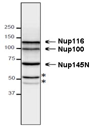
![Anti-NUP98 antibody [13C2] - BSA and Azide free (ab179894) at 0.4 µg/ml + HeLa cell extractSecondaryHRP-labeled anti-mouse IgG at 0.4 µg/mldeveloped using the ECL technique](http://www.bioprodhub.com/system/product_images/ab_products/2/sub_4/3097_ab179894-208359-ab179894wbhu.jpg)
Anti-NUP98 antibody [13C2] - BSA and Azide free (ab179894) at 0.4 µg/ml + HeLa cell extractSecondaryHRP-labeled anti-mouse IgG at 0.4 µg/mldeveloped using the ECL technique

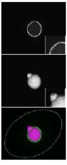
Immunofluorescent analysis of Tetrahymena thermophila cells labeling NUP98 with ab179894 at 0.5 µg/ml (top panel). DAPI (middle panel). The color image represents merged images of ab179894 (green) with DAPI (magenta).4 μg/ml of Alexa488-labelled anti-mouse IgG was used as secondary antibody. Dotted lines represent the outlines of cells. The insert is a magnified image showing the position of the micronucleus.
![All lanes : Anti-NUP98 antibody [13C2] - BSA and Azide free (ab179894)Lane 1 : Tetrahymena thermophila wild type cell extractLane 2 : Tetrahymena thermophila cell extract ectopically-expressing GFP-MacNup98A (in addition to endogenous MacNup98A)](http://www.bioprodhub.com/system/product_images/ab_products/2/sub_4/3099_ab179894-208352-ab179894wbtt.jpg)
All lanes : Anti-NUP98 antibody [13C2] - BSA and Azide free (ab179894)Lane 1 : Tetrahymena thermophila wild type cell extractLane 2 : Tetrahymena thermophila cell extract ectopically-expressing GFP-MacNup98A (in addition to endogenous MacNup98A)

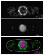
Immunofluorescent analysis of S. pombe cells labeling NUP98 with ab179894 (top panel). DAPI (middle panel). The color image represents merged images of ab179894 (green) with DAPI (magenta). Dotted lines represent the outlines of cells.
![All lanes : Anti-NUP98 antibody [13C2] - BSA and Azide free (ab179894)Lane 1 : S. pombe cell extractLane 2 : NUP98-GFP-expressing S. pombe cell extract](http://www.bioprodhub.com/system/product_images/ab_products/2/sub_4/3101_ab179894-208354-ab179894wbsp.jpg)
All lanes : Anti-NUP98 antibody [13C2] - BSA and Azide free (ab179894)Lane 1 : S. pombe cell extractLane 2 : NUP98-GFP-expressing S. pombe cell extract

Immunofluorescent analysis of S. cerevisiae cells labeling NUP98 with ab179894 at 1/100 dilution (top panel). DAPI (middle panel). The color image represents a merged image of ab179894 (green) with DAPI (magenta). Dotted lines represent the outlines of cells.

Supernatant of hybridoma culture medium of ab179894 at 1/10 dilution + S. cerevisiae cell extract

![Anti-NUP98 antibody [13C2] - BSA and Azide free (ab179894) at 0.4 µg/ml + HeLa cell extractSecondaryHRP-labeled anti-mouse IgG at 0.4 µg/mldeveloped using the ECL technique](http://www.bioprodhub.com/system/product_images/ab_products/2/sub_4/3097_ab179894-208359-ab179894wbhu.jpg)

![All lanes : Anti-NUP98 antibody [13C2] - BSA and Azide free (ab179894)Lane 1 : Tetrahymena thermophila wild type cell extractLane 2 : Tetrahymena thermophila cell extract ectopically-expressing GFP-MacNup98A (in addition to endogenous MacNup98A)](http://www.bioprodhub.com/system/product_images/ab_products/2/sub_4/3099_ab179894-208352-ab179894wbtt.jpg)

![All lanes : Anti-NUP98 antibody [13C2] - BSA and Azide free (ab179894)Lane 1 : S. pombe cell extractLane 2 : NUP98-GFP-expressing S. pombe cell extract](http://www.bioprodhub.com/system/product_images/ab_products/2/sub_4/3101_ab179894-208354-ab179894wbsp.jpg)

