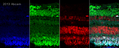
ab75882 staining OLFML2A in Human retina tissue sections by Immunohistochemistry (IHC-Fr - frozen sections). Tissue was fixed with formaldehyde and blocked with 5% milk for 12 hours at 25°C. Samples were incubated with primary antibody (1/500) for 2 hours at 25°C. A Cy3®-conjugated anti-rabbit IgG polyclonal (1/500) was used as the secondary antibody.See Abreview
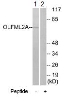
All lanes : Anti-OLFML2A antibody (ab75882) at 1/500 dilutionLane 1 : extracts from RAW264.7 cellsLane 2 : extracts from RAW264.7 cells with immunizing peptide at 10 µgLysates/proteins at 30 µg per lane.
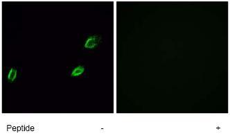
ab75882, at a dilution of 1/500-1/1000, staining OLFML2A in A549 cells by Immunofluorescence with (+) or without (-) immunising peptide.