Anti-p15 INK4b antibody
| Name | Anti-p15 INK4b antibody |
|---|---|
| Supplier | Abcam |
| Catalog | ab53034 |
| Prices | $390.00 |
| Sizes | 100 µg |
| Host | Rabbit |
| Clonality | Polyclonal |
| Isotype | IgG |
| Applications | IP WB ELISA IHC-P |
| Species Reactivities | Human, Mouse, Rat |
| Antigen | Synthetic peptide derived from human p15 INK |
| Description | Rabbit Polyclonal |
| Gene | CDKN2B |
| Conjugate | Unconjugated |
| Supplier Page | Shop |
Product images
![p15 INK4b was immunoprecipitated using 0.5mg Hela whole cell extract, 5µg of Rabbit polyclonal to p15 INK4b and 50µl of protein G magnetic beads (+). No antibody was added to the control (-).The antibody was incubated under agitation with Protein G beads for 10min, Hela whole cell extract lysate diluted in RIPA buffer was added to each sample and incubated for a further 10min under agitation.Proteins were eluted by addition of 40µl SDS loading buffer and incubated for 10min at 70°C; 10µl of each sample was separated on a SDS PAGE gel, transferred to a nitrocellulose membrane, blocked with 5% BSA and probed with ab53034.Secondary: Mouse monoclonal [SB62a] Secondary Antibody to Rabbit IgG light chain (HRP) (ab99697).Band: 15kDa; p15 INK4b](http://www.bioprodhub.com/system/product_images/ab_products/2/sub_4/5231_ab53034-196687-IPV022ab5303420mMod.jpg) p15 INK4b was immunoprecipitated using 0.5mg Hela whole cell extract, 5µg of Rabbit polyclonal to p15 INK4b and 50µl of protein G magnetic beads (+). No antibody was added to the control (-).The antibody was incubated under agitation with Protein G beads for 10min, Hela whole cell extract lysate diluted in RIPA buffer was added to each sample and incubated for a further 10min under agitation.Proteins were eluted by addition of 40µl SDS loading buffer and incubated for 10min at 70°C; 10µl of each sample was separated on a SDS PAGE gel, transferred to a nitrocellulose membrane, blocked with 5% BSA and probed with ab53034.Secondary: Mouse monoclonal [SB62a] Secondary Antibody to Rabbit IgG light chain (HRP) (ab99697).Band: 15kDa; p15 INK4b
p15 INK4b was immunoprecipitated using 0.5mg Hela whole cell extract, 5µg of Rabbit polyclonal to p15 INK4b and 50µl of protein G magnetic beads (+). No antibody was added to the control (-).The antibody was incubated under agitation with Protein G beads for 10min, Hela whole cell extract lysate diluted in RIPA buffer was added to each sample and incubated for a further 10min under agitation.Proteins were eluted by addition of 40µl SDS loading buffer and incubated for 10min at 70°C; 10µl of each sample was separated on a SDS PAGE gel, transferred to a nitrocellulose membrane, blocked with 5% BSA and probed with ab53034.Secondary: Mouse monoclonal [SB62a] Secondary Antibody to Rabbit IgG light chain (HRP) (ab99697).Band: 15kDa; p15 INK4b
 All lanes : Anti-p15 INK4b antibody (ab53034) at 1/500 dilutionLane 1 : HeLa cell extractLane 2 : HeLa cell extract with peptide
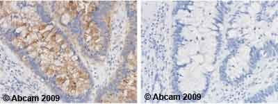 Left panel: with primary antibody at 2 ug/ml. Right panel: isotype control. Sections were stained using an automated system (Dako PT Link), at room temperature. Sections were rehydrated and antigen retrieved with the Dako 3-in-1 AR buffers EDTA pH 9.0. Slides were peroxidase blocked in 3% H2O2 in methanol for 10 minutes. They were then blocked with Dako Protein block for 10 minutes (containing casein 0.25% in PBS) then incubated with primary antibody for 20 minutes and detected with Dako Envision Flex amplification kit for 30 minutes. Colorimetric detection was completed with diaminobenzidine for 5 minutes. Slides were counterstained with Haematoxylin and coverslipped under DePeX. Please note that for manual staining we recommend to optimize the primary antibody concentration and incubation time (overnight incubation), and amplification may be required.
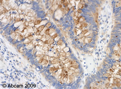 ab53034 (2µg/ml) staining p15 INK4b in human colon carcinoma.Sections were stained using an automated system (DAKO Autostainer Plus ), at room temperature: sections were rehydrated and antigen retrieved with the Dako 3 in 1 AR buffers EDTA pH 9.0 . Slides were peroxidase blocked in 3% H2O2 in methanol for 10 mins. They were then blocked with Dako Protein block for 10 minutes (containing casein 0.25% in PBS) then incubated with primary antibody for 20 min and detected with Dako envision flex amplification kit for 30 minutes. Colorimetric detection was completed with Diaminobenzidine for 5 minutes. Slides were counterstained with Haematoxylin and coverslipped under DePeX. Please note that for manual staining we recommend to optimize the primary antibody concentration and incubation time (overnight incubation), and amplification may be required.
Product References
A KRAS-directed transcriptional silencing pathway that mediates the CpG island - A KRAS-directed transcriptional silencing pathway that mediates the CpG island
Serra RW, Fang M, Park SM, Hutchinson L, Green MR. Elife. 2014 Mar 12;3:e02313.
Functional analyses of coronary artery disease associated variation on chromosome - Functional analyses of coronary artery disease associated variation on chromosome
Motterle A, Pu X, Wood H, Xiao Q, Gor S, Ng FL, Chan K, Cross F, Shohreh B, Poston RN, Tucker AT, Caulfield MJ, Ye S. Hum Mol Genet. 2012 Sep 15;21(18):4021-9.
Estradiol partially recapitulates murine pituitary cell cycle response to - Estradiol partially recapitulates murine pituitary cell cycle response to
Toledano Y, Zonis S, Ren SG, Wawrowsky K, Chesnokova V, Melmed S. Endocrinology. 2012 Oct;153(10):5011-22. Epub 2012 Jul 31.