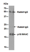
Western blot analysis of immunoprecipitation pellet from HeLa lysate immunoprecipitated using ab192239 at 1/30 dilution.Secondary: Goat Anti-Rabbit IgG, (H+L), Peroxidase conjugate at 1/1000 dilution.
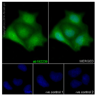
Immunofluorescent analysis of HeLa cells (4% Paraformaldehyde-fixed, 0.1% tritonX-100 permeabilized) labeling p18 INK4C with ab192239 at 1/100 dilution (5μg/mL) followed by Goat anti rabbit IgG (AlexaFluor® 488) (ab150077) secondary at 1/200 dilution and counter-stained with DAPI (blue).Negative controls: anti-p18 INK4C at 1/100 dilution, Secondary ab (Goat anti mouse IgG (Alexa Fluor®594)) at 1/400 dilution.
Immunohistochemical analysis of paraffin-embedded Human glioma tissue labeling p18 INK4C with ab192239 at 1/50 dilution followed by pre-diluted HRP Polymer for Rabbit/Mouse IgG secondary antibody and counter-stained with Hematoxylin. (inset: negative control).
Immunohistochemical analysis of paraffin-embedded Human brain tissue labeling p18 INK4C with ab192239 at 1/50 dilution followed by pre-diluted HRP Polymer for Rabbit/Mouse IgG secondary antibody and counter-stained with Hematoxylin. (inset: negative control).
![All lanes : Anti-p18 INK4c antibody [EPR15891] (ab192239) at 1/1000 dilutionLane 1 : Rat kidney lysateLane 2 : Rat spleen lysateLane 3 : NIH 3T3 lysateLysates/proteins at 10 µg per lane.SecondaryGoat Anti-Rabbit IgG, (H+L), Peroxidase conjugate at 1/1000 dilution](http://www.bioprodhub.com/system/product_images/ab_products/2/sub_4/5293_ab192239-230025-ab192239b.jpg)
All lanes : Anti-p18 INK4c antibody [EPR15891] (ab192239) at 1/1000 dilutionLane 1 : Rat kidney lysateLane 2 : Rat spleen lysateLane 3 : NIH 3T3 lysateLysates/proteins at 10 µg per lane.SecondaryGoat Anti-Rabbit IgG, (H+L), Peroxidase conjugate at 1/1000 dilution
![All lanes : Anti-p18 INK4c antibody [EPR15891] (ab192239) at 1/10000 dilutionLane 1 : 293 cell lysateLane 2 : Ramos cell lysateLane 3 : HeLa cell lysateLysates/proteins at 20 µg per lane.SecondaryGoat Anti-Rabbit IgG, (H+L), Peroxidase conjugate at 1/1000 dilution](http://www.bioprodhub.com/system/product_images/ab_products/2/sub_4/5294_ab192239-230024-ab192239.jpg)
All lanes : Anti-p18 INK4c antibody [EPR15891] (ab192239) at 1/10000 dilutionLane 1 : 293 cell lysateLane 2 : Ramos cell lysateLane 3 : HeLa cell lysateLysates/proteins at 20 µg per lane.SecondaryGoat Anti-Rabbit IgG, (H+L), Peroxidase conjugate at 1/1000 dilution