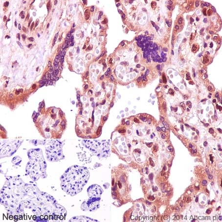
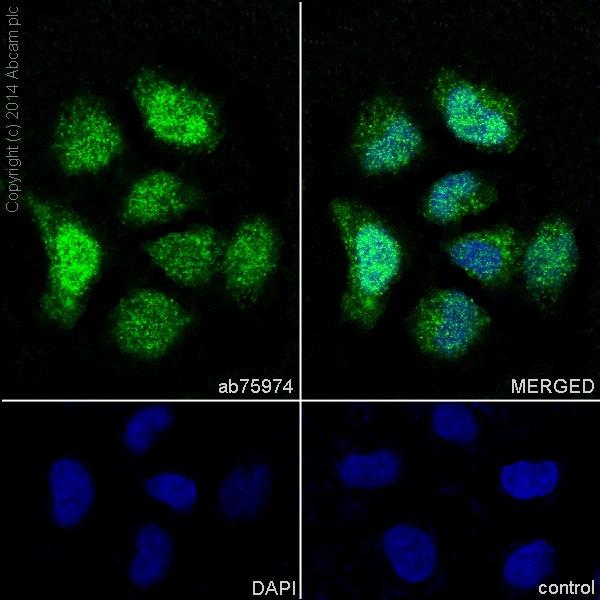
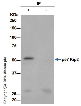
Anti-p57 Kip2 antibody [EP2515Y]
| Name | Anti-p57 Kip2 antibody [EP2515Y] |
|---|---|
| Supplier | Abcam |
| Catalog | ab75974 |
| Prices | $392.00 |
| Sizes | 100 µl |
| Host | Rabbit |
| Clonality | Monoclonal |
| Isotype | IgG |
| Clone | EP2515Y |
| Applications | ICC/IF ICC/IF WB IP IHC-P |
| Species Reactivities | Mouse, Rat, Human |
| Antigen | Synthetic peptide (the amino acid sequence is considered to be commercially sensitive) corresponding to Human p57 Kip2 aa 50-150 (N terminal) |
| Description | Rabbit Monoclonal |
| Gene | CDKN1C |
| Conjugate | Unconjugated |
| Supplier Page | Shop |
Product images
Product References
Frs2alpha and Shp2 signal independently of Gab to mediate FGF signaling in lens - Frs2alpha and Shp2 signal independently of Gab to mediate FGF signaling in lens
Li H, Tao C, Cai Z, Hertzler-Schaefer K, Collins TN, Wang F, Feng GS, Gotoh N, Zhang X. J Cell Sci. 2014 Feb 1;127(Pt 3):571-82.
Liver X Receptor activation delays chondrocyte hypertrophy during endochondral - Liver X Receptor activation delays chondrocyte hypertrophy during endochondral
Sun MM, Beier F. Osteoarthritis Cartilage. 2014 Jul;22(7):996-1006. doi:
Histone deacetylase 2 (HDAC2) regulates chromosome segregation and kinetochore - Histone deacetylase 2 (HDAC2) regulates chromosome segregation and kinetochore
Ma P, Schultz RM. PLoS Genet. 2013;9(3):e1003377.
CDKN1C mutation affecting the PCNA-binding domain as a cause of familial Russell - CDKN1C mutation affecting the PCNA-binding domain as a cause of familial Russell
Brioude F, Oliver-Petit I, Blaise A, Praz F, Rossignol S, Le Jule M, Thibaud N, Faussat AM, Tauber M, Le Bouc Y, Netchine I. J Med Genet. 2013 Dec;50(12):823-30.
Clonal-level responses of functionally distinct hematopoietic stem cells to - Clonal-level responses of functionally distinct hematopoietic stem cells to
Mallaney C, Kothari A, Martens A, Challen GA. Exp Hematol. 2014 Apr;42(4):317-327.e2.
PR-domain-containing Mds1-Evi1 is critical for long-term hematopoietic stem cell - PR-domain-containing Mds1-Evi1 is critical for long-term hematopoietic stem cell
Zhang Y, Stehling-Sun S, Lezon-Geyda K, Juneja SC, Coillard L, Chatterjee G, Wuertzer CA, Camargo F, Perkins AS. Blood. 2011 Oct 6;118(14):3853-61.
Checkpoint kinase 1 prevents cell cycle exit linked to terminal cell - Checkpoint kinase 1 prevents cell cycle exit linked to terminal cell
Ullah Z, de Renty C, DePamphilis ML. Mol Cell Biol. 2011 Oct;31(19):4129-43.
![Anti-p57 Kip2 antibody [EP2515Y] (ab75974) at 1/500 dilution (purified) + Human brain at 20 µgSecondaryHRP-conjugated anti-rabbit IgG, specific to the non-reduced form of IgG at 1/1000 dilution](http://www.bioprodhub.com/system/product_images/ab_products/2/sub_4/6004_ab75974-237204-ab75974wb1.jpg)
![All lanes : Anti-p57 Kip2 antibody [EP2515Y] (ab75974) at 1/500 dilution (purified)Lane 1 : Mouse brainLane 2 : Rat brainLysates/proteins at 10 µg per lane.SecondaryPeroxidase-conjugated goat anti-rabbit IgG (H+L) at 1/1000 dilution](http://www.bioprodhub.com/system/product_images/ab_products/2/sub_4/6005_ab75974-237203-ab75974wb2.jpg)
![All lanes : Anti-p57 Kip2 antibody [EP2515Y] (ab75974) at 1/500 dilution (unpurified)Lane 1 : HeLa lysateLane 2 : Jurkat lysateLane 3 : Human brain lysateLysates/proteins at 10 µg per lane.Secondarygoat anti-rabbit HRP at 1/2000 dilution](http://www.bioprodhub.com/system/product_images/ab_products/2/sub_4/6006_ab75974wb.gif)


