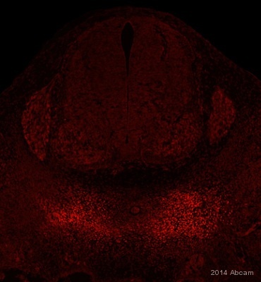
ab95227 staining PAX1 in mouse E11.5 embryo tissue sections by Immunohistochemistry (IHC-Fr - frozen sections). Tissue was fixed with 2% paraformaldehyde in PBS for 2 hours at 4°C then placed in 30% sucrose overnight at 4°C prior to embedding in OCT using liquid nitrogen. Tissue was blocked with 1X PBS + 1.5% Milk + 1.5% BSA + 0.1% Triton X-100 for 12 hours at 4°C. Samples were incubated with primary antibody (1/500 in blocking buffer) for 1 hour at 23°C. An Alexa Fluor® 568-conjugated goat anti-rabbit IgG polyclonal (1/250) was used as the secondary antibody.See Abreview

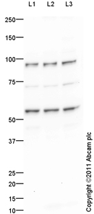
All lanes : Anti-PAX1 antibody (ab95227) at 1 µg/mlLane 1 : Jurkat (Human T cell lymphoblast-like cell line) Whole Cell LysateLane 2 : Raji (Human Burkitt's lymphoma cell line) Whole Cell Lysate Lane 3 : K562 (Human erythromyeloblastoid leukemia cell line) Whole Cell Lysate Lysates/proteins at 10 µg per lane.SecondaryGoat Anti-Rabbit IgG H&L (HRP) preadsorbed (ab97080) at 1/5000 dilutiondeveloped using the ECL techniquePerformed under reducing conditions.

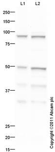
All lanes : Anti-PAX1 antibody (ab95227) at 1 µg/mlLane 1 : Thymus (Mouse) Tissue Lysate Lane 2 : Thymus (Rat) Tissue Lysate Lysates/proteins at 10 µg per lane.SecondaryGoat Anti-Rabbit IgG H&L (HRP) preadsorbed (ab97080) at 1/5000 dilutiondeveloped using the ECL techniquePerformed under reducing conditions.


