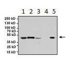
All lanes : Anti-PAX5 antibody (ab183575) at 1/1000 dilutionLane 1 : Ramos whole cell lysateLane 2 : Daudi whole cell lysateLane 3 : Raji whole cell lysateLane 4 : Jurkat whole cell lysateLane 5 : A20 whole cell lysateLysates/proteins at 75 µg per lane.SecondaryGoat anti rabbit HRP at 1/500 dilution

Immunofluorescent analysis of PAX5 (green) in Ramos and negative control Jurkat cells. Formalin fixed cells were permeabilized with 0.1% Triton X-100 in TBS for 10 minutes at room temperature. Cells were blocked with 2% Blocker BSA for 15 minutes at room temperature. Cells were probed with ab183575 at a dilution of 1/100 for at least 1 hour at room temperature, washed with PBS, and incubated with a DyLight 488-conjugated goat anti-rabbit IgG secondary antibody at a dilution of 1/200 for 30 minutes at room temperature. F-Actin (red) was stained with DyLight-554 Phalloidin and nuclei (blue) were stained with Hoechst 33342 dye.

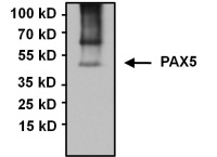
Immunoprecipitation of PAX5 was performed using Raji whole cell lysates. Antigen-antibody complexes were formed by incubating 75 ug of lysate with 5ug of ab183575 overnight on a rocking platform at 4°C. The immune complexes were captured on 50ul Protein A/G Agarose, washed extensively, and eluted with 5X Lane Marker Reducing Sample Buffer. Samples was resolved on a 4-20% Tris-HCl polyacrylamide gel, transferred to a PVDF membrane, and blocked with 5% BSA/TBS-0.1%Tween-20 for 1 hour. The membrane was probed with ab183575 at a dilution of 1/1000 overnight rotating at 4°C. Membranes were washed in TBST, and probed with an IP Detection Reagent at a dilution of 1/1000 for at least one hour. Chemiluminescent detection was performed.

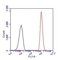
Flow cytometry analysis of PAX5 (red histogram) on Ramos cells. Cells were harvested, fixed with 4% formaldehyde, permeabilized, washed with PBS, and incubated with ab183575 at a 1/50 dilution or with PBS alone (black histogram) for 1 hour at room temperature. For flow cytometry analysis, 30-minute incubation with DyLight 488 goat anti-rabbit IgG secondary antibody was performed. 30,000 cells were acquired for analysis.


