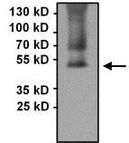
Detection of PAX8 in Immunoprecipitates of COS7 whole cell lysates. Antigen-antibody complexes were formed by incubating 500μg of lysate with 3μg of ab183578. The membrane was probed with ab183578 at 1/1000 dilution.
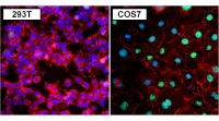
Immunofluorescent analysis of 293T (left) and COS7 (right) cells (formalin-fixed, 0.1% Triton X-100 permeabilized) labeling PAX8 with ab183578 at 1/400 dilution followed by DyLight 488 goat anti-rabbit IgG secondary antibody at 1/400 dilution. Nuclei (blue) were stained with Hoechst 33342 dye.
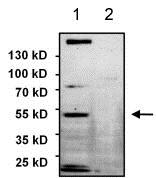
All lanes : Anti-PAX8 antibody (ab183578) at 1/1000 dilutionLane 1 : SKOV3 whole cell lysateLane 2 : HeLa whole cell lysateLysates/proteins at 75 µg per lane.SecondaryGoat anti-rabbit HRP at 1/500 dilutiondeveloped using the ECL technique
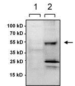
All lanes : Anti-PAX8 antibody (ab183578) at 1/1000 dilutionLane 1 : HEK293 empty vector controlLane 2 : HEK293 lysate overexpressing PAX8Lysates/proteins at 40 µg per lane.SecondaryGoat anti-rabbit HRP at 1/500 dilutiondeveloped using the ECL technique
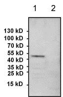
All lanes : Anti-PAX8 antibody (ab183578) at 1/1000 dilutionLane 1 : COS7 whole cell lysateLane 2 : HeLa whole cell lysateLysates/proteins at 60 µg per lane.SecondaryGoat anti-rabbit HRP at 1/500 dilutiondeveloped using the ECL technique


