![All lanes : Anti-PAX8 antibody [1F8-3A8] (ab183573) at 1/1000 dilutionLane 1 : SKOV-3 cell lysateLane 2 : COS7 cell lysateLane 3 : Negative control HeLa whole cell lysatesLysates/proteins at 60 µg per lane.SecondaryHRP-conjugated goat anti-mouse IgG at 1/500 dilution](http://www.bioprodhub.com/system/product_images/ab_products/2/sub_4/7818_ab183573-213841-ab1835731.jpg)
All lanes : Anti-PAX8 antibody [1F8-3A8] (ab183573) at 1/1000 dilutionLane 1 : SKOV-3 cell lysateLane 2 : COS7 cell lysateLane 3 : Negative control HeLa whole cell lysatesLysates/proteins at 60 µg per lane.SecondaryHRP-conjugated goat anti-mouse IgG at 1/500 dilution

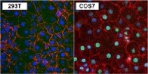
Immunofluorescent analysis of PAX8 (green) in 293T (left panel) and COS7 cells (right panel) using ab183573 at a 1/50 dilution. Formalin fixed cells were permeabilized with 0.1% Triton X-100 in TBS for 10 minutes at room temperature. Cells were blocked with 1% blocker BSA for 15 minutes at room temperature. A DyLight 488-conjugated goat anti-mouse IgG secondary antibody was used at a dilution of 1/400 for 30 minutes at room temperature. F-Actin (red) was stained with DyLight-554 Phalloidin and nuclei (blue) were stained with Hoechst 33342 dye.
![All lanes : Anti-PAX8 antibody [1F8-3A8] (ab183573) at 1/1000 dilutionLane 1 : untransfected HEK293T cell lysateLane 2 : HEK293T cell lysate over expressing PAX8Lysates/proteins at 25 µg per lane.SecondaryHRP-conjugated goat anti-mouse IgG at 1/500 dilution](http://www.bioprodhub.com/system/product_images/ab_products/2/sub_4/7820_ab183573-213840-ab1835732.jpg)
All lanes : Anti-PAX8 antibody [1F8-3A8] (ab183573) at 1/1000 dilutionLane 1 : untransfected HEK293T cell lysateLane 2 : HEK293T cell lysate over expressing PAX8Lysates/proteins at 25 µg per lane.SecondaryHRP-conjugated goat anti-mouse IgG at 1/500 dilution
![All lanes : Anti-PAX8 antibody [1F8-3A8] (ab183573) at 1/1000 dilutionLane 1 : SKOV-3 cell lysateLane 2 : COS7 cell lysateLane 3 : Negative control HeLa whole cell lysatesLysates/proteins at 60 µg per lane.SecondaryHRP-conjugated goat anti-mouse IgG at 1/500 dilution](http://www.bioprodhub.com/system/product_images/ab_products/2/sub_4/7818_ab183573-213841-ab1835731.jpg)

![All lanes : Anti-PAX8 antibody [1F8-3A8] (ab183573) at 1/1000 dilutionLane 1 : untransfected HEK293T cell lysateLane 2 : HEK293T cell lysate over expressing PAX8Lysates/proteins at 25 µg per lane.SecondaryHRP-conjugated goat anti-mouse IgG at 1/500 dilution](http://www.bioprodhub.com/system/product_images/ab_products/2/sub_4/7820_ab183573-213840-ab1835732.jpg)