![All lanes : Anti-PDLIM2 antibody [9B6] (ab119093) at 1/500 dilutionLane 1 : Lysate from HEK293T cells transfected with vector alone.Lane 2 : Lysate from HEK293T cells transfected with a vector expressing full length PDLIM2.Lysates/proteins at 5 µg per lane.developed using the ECL technique](http://www.bioprodhub.com/system/product_images/ab_products/2/sub_4/9398_PDLIM2-Primary-antibodies-ab119093-1.jpg)
All lanes : Anti-PDLIM2 antibody [9B6] (ab119093) at 1/500 dilutionLane 1 : Lysate from HEK293T cells transfected with vector alone.Lane 2 : Lysate from HEK293T cells transfected with a vector expressing full length PDLIM2.Lysates/proteins at 5 µg per lane.developed using the ECL technique
![All lanes : Anti-PDLIM2 antibody [9B6] (ab119093) at 1/500 dilutionLane 1 : Extracts from HepG2 cell line.Lane 2 : Extracts from HeLa cell line.Lane 3 : Extracts from SVT2 cell line.Lane 4 : Extracts from A549 cell line.Lane 5 : Extracts from COS7 cell line.Lane 6 : Extracts from Jurkat cell line.Lane 7 : Extracts from MDCK cell line.Lane 8 : Extracts from PC12 cell line.Lane 9 : Extracts from MCF7 cell line.Lysates/proteins at 35 µg per lane.developed using the ECL technique](http://www.bioprodhub.com/system/product_images/ab_products/2/sub_4/9399_PDLIM2-Primary-antibodies-ab119093-2.JPG)
All lanes : Anti-PDLIM2 antibody [9B6] (ab119093) at 1/500 dilutionLane 1 : Extracts from HepG2 cell line.Lane 2 : Extracts from HeLa cell line.Lane 3 : Extracts from SVT2 cell line.Lane 4 : Extracts from A549 cell line.Lane 5 : Extracts from COS7 cell line.Lane 6 : Extracts from Jurkat cell line.Lane 7 : Extracts from MDCK cell line.Lane 8 : Extracts from PC12 cell line.Lane 9 : Extracts from MCF7 cell line.Lysates/proteins at 35 µg per lane.developed using the ECL technique
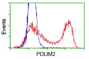
ab119093 at 1/100 dilution staining PDLIM2 in HEK293T cells transfected with either a vector expressing PDLIM2 (Red) or an empty vector control plasmid (Blue) and then analysed by Flow Cytometry.
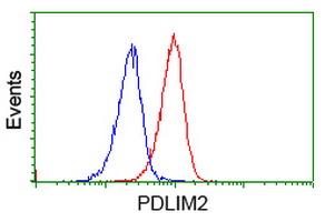
Flow cytometric Analysis of Jurkat cells, using ab119093 at 1/100 dilution (Red), compared to using a nonspecific negative control antibody(Blue).