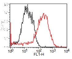
Overlay histogram showing HUVEC cells stained with ab125007 (red line) at 1/40 dilution. The cells were fixed with 2% paraformaldehyde. The secondary antibody used was a FITC conjugated goat anti-rabbit IgG at 1/150 dilution. Isotype control antibody (black line) was rabbit monoclonal IgG used under the same conditions.
![Anti-Pentraxin 3 antibody [EPR6699] (ab125007) at 1/10000 dilution + NIH/3T3 cell lysate at 20 µgSecondaryGoat Anti-Rabbit IgG, (H+L), HRP-conjugated at 1/1000 dilution](http://www.bioprodhub.com/system/product_images/ab_products/2/sub_4/9766_ab125007-240399-125007-WB2.jpg)
Anti-Pentraxin 3 antibody [EPR6699] (ab125007) at 1/10000 dilution + NIH/3T3 cell lysate at 20 µgSecondaryGoat Anti-Rabbit IgG, (H+L), HRP-conjugated at 1/1000 dilution
![Anti-Pentraxin 3 antibody [EPR6699] (ab125007) at 1/10000 dilution + HUVEC cell lysate at 20 µgSecondaryGoat Anti-Rabbit IgG, (H+L), HRP-conjugated at 1/1000 dilution](http://www.bioprodhub.com/system/product_images/ab_products/2/sub_4/9767_ab125007-240395-125007-WB1.jpg)
Anti-Pentraxin 3 antibody [EPR6699] (ab125007) at 1/10000 dilution + HUVEC cell lysate at 20 µgSecondaryGoat Anti-Rabbit IgG, (H+L), HRP-conjugated at 1/1000 dilution
![All lanes : Anti-Pentraxin 3 antibody [EPR6699] (ab125007) at 1/1000 dilution (unpurified)Lane 1 : HL-60 treated with Dexamethasone lysateLane 2 : HL-60 lysateLane 3 : Human spleen lysateLane 4 : NIH/3T3 cell lysateLane 5 : HUVEC lysateLysates/proteins at 10 µg per lane.SecondaryHRP labelled goat anti-rabbit at 1/2000 dilution](http://www.bioprodhub.com/system/product_images/ab_products/2/sub_4/9768_Pentraxin-3-Primary-antibodies-ab125007-1.jpg)
All lanes : Anti-Pentraxin 3 antibody [EPR6699] (ab125007) at 1/1000 dilution (unpurified)Lane 1 : HL-60 treated with Dexamethasone lysateLane 2 : HL-60 lysateLane 3 : Human spleen lysateLane 4 : NIH/3T3 cell lysateLane 5 : HUVEC lysateLysates/proteins at 10 µg per lane.SecondaryHRP labelled goat anti-rabbit at 1/2000 dilution

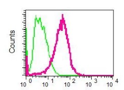
Flow cytometric analysis of permeabilized HUVEC cells using ab125007, unpurified, at 1/10 dilution (red) or a rabbit IgG (negative) (green).

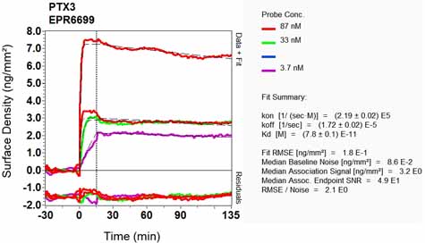
Equilibrium disassociation constant (KD)Learn more about KD Click here to learn more about KD

![Anti-Pentraxin 3 antibody [EPR6699] (ab125007) at 1/10000 dilution + NIH/3T3 cell lysate at 20 µgSecondaryGoat Anti-Rabbit IgG, (H+L), HRP-conjugated at 1/1000 dilution](http://www.bioprodhub.com/system/product_images/ab_products/2/sub_4/9766_ab125007-240399-125007-WB2.jpg)
![Anti-Pentraxin 3 antibody [EPR6699] (ab125007) at 1/10000 dilution + HUVEC cell lysate at 20 µgSecondaryGoat Anti-Rabbit IgG, (H+L), HRP-conjugated at 1/1000 dilution](http://www.bioprodhub.com/system/product_images/ab_products/2/sub_4/9767_ab125007-240395-125007-WB1.jpg)
![All lanes : Anti-Pentraxin 3 antibody [EPR6699] (ab125007) at 1/1000 dilution (unpurified)Lane 1 : HL-60 treated with Dexamethasone lysateLane 2 : HL-60 lysateLane 3 : Human spleen lysateLane 4 : NIH/3T3 cell lysateLane 5 : HUVEC lysateLysates/proteins at 10 µg per lane.SecondaryHRP labelled goat anti-rabbit at 1/2000 dilution](http://www.bioprodhub.com/system/product_images/ab_products/2/sub_4/9768_Pentraxin-3-Primary-antibodies-ab125007-1.jpg)

