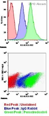
Anti-Peroxiredoxin 6 antibody
| Name | Anti-Peroxiredoxin 6 antibody |
|---|---|
| Supplier | Abcam |
| Catalog | ab59543 |
| Prices | $403.00 |
| Sizes | 100 µl |
| Host | Rabbit |
| Clonality | Polyclonal |
| Isotype | IgG |
| Applications | FC WB ELISA IHC-F IHC-P ICC/IF ICC/IF |
| Species Reactivities | Mouse, Rat, Human |
| Antigen | Recombinant full length protein (Rat) Peroxiredoxin 6 |
| Description | Rabbit Polyclonal |
| Gene | PRDX6 |
| Conjugate | Unconjugated |
| Supplier Page | Shop |
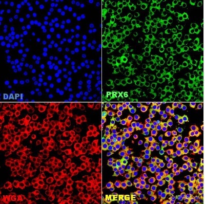
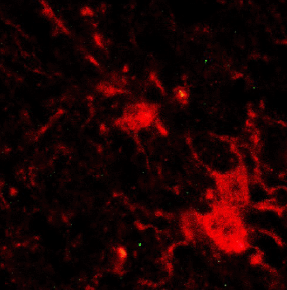
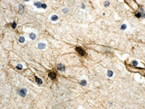
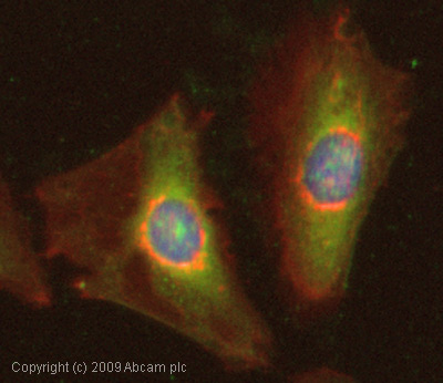
Product images
Product References
Impact of genomic stability on protein expression in endometrioid endometrial - Impact of genomic stability on protein expression in endometrioid endometrial
Lomnytska MI, Becker S, Gemoll T, Lundgren C, Habermann J, Olsson A, Bodin I, Engstrom U, Hellman U, Hellman K, Hellstrom AC, Andersson S, Mints M, Auer G. Br J Cancer. 2012 Mar 27;106(7):1297-305.
Synaptic protein expression is regulated by a pro-oxidant diet in APPxPS1 mice. - Synaptic protein expression is regulated by a pro-oxidant diet in APPxPS1 mice.
Broadstock M, Lewinsky R, Jones EL, Mitchelmore C, Howlett DR, Francis PT. J Neural Transm. 2012 Apr;119(4):493-6.
In vivo matrix metalloproteinase-7 substrates identified in the left ventricle - In vivo matrix metalloproteinase-7 substrates identified in the left ventricle
Chiao YA, Zamilpa R, Lopez EF, Dai Q, Escobar GP, Hakala K, Weintraub ST, Lindsey ML. J Proteome Res. 2010 May 7;9(5):2649-57.
Protection of peroxiredoxin II on oxidative stress-induced cardiomyocyte death - Protection of peroxiredoxin II on oxidative stress-induced cardiomyocyte death
Zhao W, Fan GC, Zhang ZG, Bandyopadhyay A, Zhou X, Kranias EG. Basic Res Cardiol. 2009 Jul;104(4):377-89.
