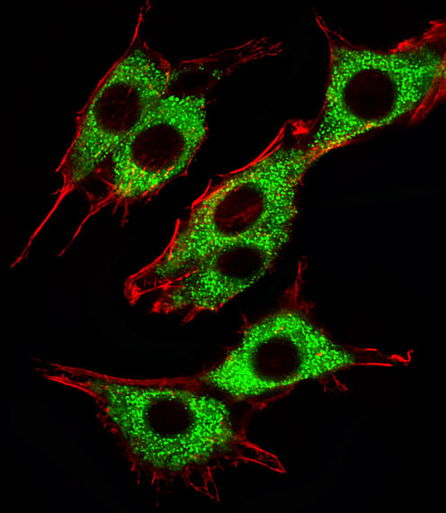
Immunocytochemistry/Immunofluorescence analysis of PC-12 cells labelling PINK1 (green) with ab75487 at a dilution of 1/25. An Alexa Fluor® 488-conjugated goat anti-mouse IgG was used as the secondary antibody (1/400). Cytoplasmic actin was counterstained with Alexa Fluor® 555-conjugated with Phalloidin (red).
![All lanes : Anti-PINK1 antibody [38CT18.7] (ab75487) at 1/1000 dilutionLane 1 : A431 cell lysateLane 2 : Mouse brain tissue lysateLysates/proteins at 20 µg per lane.SecondaryHRP-conjugated goat anti-rabbit IgG (H+L) at 1/5000 dilution](http://www.bioprodhub.com/system/product_images/ab_products/2/sub_4/12083_ab75487-239457-ab75487wb.jpg)
All lanes : Anti-PINK1 antibody [38CT18.7] (ab75487) at 1/1000 dilutionLane 1 : A431 cell lysateLane 2 : Mouse brain tissue lysateLysates/proteins at 20 µg per lane.SecondaryHRP-conjugated goat anti-rabbit IgG (H+L) at 1/5000 dilution
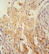
Immunohistochemistry (Formalin/PFA-fixed paraffin-embedded sections) analysis of human kidney tissue labelling PINK1 with ab75487. A peroxidase-conjugated anti-mouse IgG was used as the secondary antibody, followed by DAB staining.
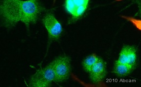
Immunocytochemistry/Immunofluorescence analysis of PC12 cells labelling PINK1 with ab75487. Cells were fixed with paraformaldehyde, permeabilization with 0.3X Triton X-100 and blocked with 10% serum for 1 hour at 4°C. Cells were incubated with the primary antibody at 2 µg/ml for 24 hours at 4°C. An Alexa Fluor® 488-conjugated anti-mouse (1/1000) was used as the secondary antibody. DAPI was used to stain the cell nuclei (blue) at a concentration of 1.43µM.See Abreview