![All lanes : Anti-PPM1G antibody [EPR13362-31] (ab186423) at 1/10000 dilutionLane 1 : 293T cell lysateLane 2 : Jurkat cell lysateLane 3 : HeLa cell lysateLane 4 : MCF7 cell lysateLysates/proteins at 20 µg per lane.SecondaryGoat Anti-Rabbit IgG, (H+L), Peroxidase conjugate at 1/1000 dilution](http://www.bioprodhub.com/system/product_images/ab_products/2/sub_4/15006_ab186423-219982-ab186423WB.jpg)
All lanes : Anti-PPM1G antibody [EPR13362-31] (ab186423) at 1/10000 dilutionLane 1 : 293T cell lysateLane 2 : Jurkat cell lysateLane 3 : HeLa cell lysateLane 4 : MCF7 cell lysateLysates/proteins at 20 µg per lane.SecondaryGoat Anti-Rabbit IgG, (H+L), Peroxidase conjugate at 1/1000 dilution
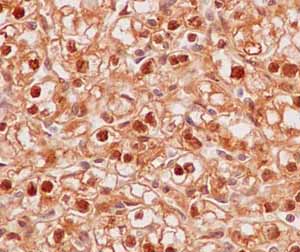
Immunohistochemical analysis of paraffin-embedded Human kidney clear cell carcinoma tissue labeling PPM1G with ab186423 at 1/500 dilution followed by prediluted HRP Polymer for Rabbit IgG. Counter stained with Hematoxylin.
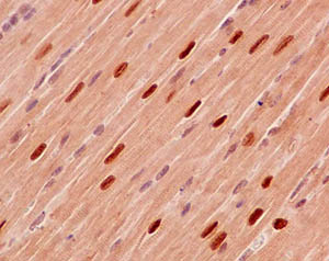
Immunohistochemical analysis of paraffin-embedded Mouse cardiac muscle tissue labeling PPM1G with ab186423 at 1/500 dilution followed by prediluted HRP Polymer for Rabbit IgG. Counter stained with Hematoxylin.
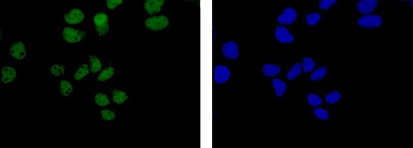
Immunofluorescent analysis of 4% paraformaldehyde-fixed HeLa cells labeling PPM1G with ab186423 at 1/500 dilution followed by Goat anti rabbit IgG (Alexa Fluor® 488) secondary antibody at 1/200 dilution (Green). Counter stained with Dapi (Blue).
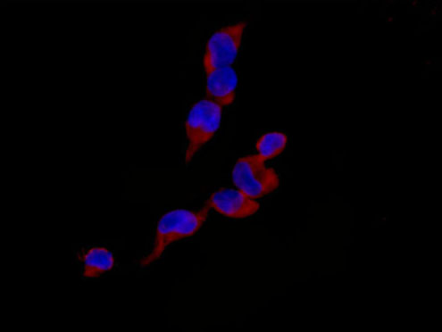
Immunofluorescent analysis of 4% paraformaldehyde-fixed 293T cells labeling PPM1G with ab186423 at 1/100 dilution followed by Goat anti rabbit IgG (Alexa Fluor® 555) secondary antibody at 1/200 dilution. Counter stained with Dapi.
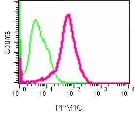
Flow cytometric analysis of 2% paraformaldehyde-fixed 293 cells labeling PPM1G with ab186423 at 1/200 dilution (red) compared to a Rabbit monoclonal IgG isotype control (green), followed by Goat anti rabbit IgG (FITC) secondary antibody at 1/150 dilution.