![All lanes : Anti-PRDM4/PFM1 antibody [EPR9432] (ab156867) at 1/1000 dilutionLane 1 : HeLa cell lysateLane 2 : 293 cell lysateLane 3 : PC-3 cell lysateLane 4 : SH-SY5Y cell lysateLysates/proteins at 20 µg per lane.SecondaryPeroxidase-conjugated goat anti-rabbit IgG (H+L) at 1/1000 dilution](http://www.bioprodhub.com/system/product_images/ab_products/2/sub_4/15640_ab156867-220968-ab156867wb.jpg)
All lanes : Anti-PRDM4/PFM1 antibody [EPR9432] (ab156867) at 1/1000 dilutionLane 1 : HeLa cell lysateLane 2 : 293 cell lysateLane 3 : PC-3 cell lysateLane 4 : SH-SY5Y cell lysateLysates/proteins at 20 µg per lane.SecondaryPeroxidase-conjugated goat anti-rabbit IgG (H+L) at 1/1000 dilution
![All lanes : Anti-PRDM4/PFM1 antibody [EPR9432] (ab156867) at 1/1000 dilutionLane 1 : Mouse heart tissue lysateLane 2 : Rat heart tissue lysateLysates/proteins at 10 µg per lane.SecondaryPeroxidase-conjugated goat anti-rabbit IgG (H + L) at 1/1000 dilution](http://www.bioprodhub.com/system/product_images/ab_products/2/sub_4/15641_ab156867-220969-ab156867wb2.jpg)
All lanes : Anti-PRDM4/PFM1 antibody [EPR9432] (ab156867) at 1/1000 dilutionLane 1 : Mouse heart tissue lysateLane 2 : Rat heart tissue lysateLysates/proteins at 10 µg per lane.SecondaryPeroxidase-conjugated goat anti-rabbit IgG (H + L) at 1/1000 dilution

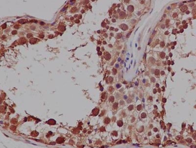
Immunohistochemistry (Formalin/PFA-fixed paraffin-embedded sections) analysis of Human testis tissue labelling PRDM1/PFM1 with ab156867 at 1/500. An ImmunoHistoprobe (Ready to use) HRP polymer for Rabbit IgG was used as the secondary antibody. Tissue was counterstained with Hematoxylin.

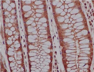
Immunohistochemistry (Formalin/PFA-fixed paraffin-embedded sections) analysis of Human colon tissue labelling PRDM1/PFM1 with ab156867 at 1/500. An ImmunoHistoprobe (Ready to use) HRP polymer for Rabbit IgG was used as the secondary antibody. Tissue was counterstained with Hematoxylin.

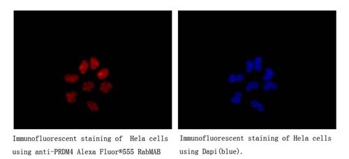
Immunocytochemistry/Immunofluorescence analysis of HeLa cells labelling PRDM4/PFM1 with ab156867 at 1/250. Cells were fixed with 4% paraformaldehyde. An Alexa Fluor® 555-conjugtaed goat anti-rabbit IgG (1/200) was used as the secondary antibody. Counterstained with DAPI.

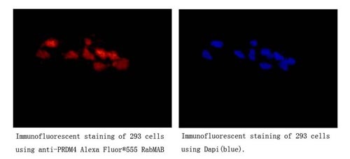
Immunocytochemistry/Immunofluorescence analysis of 293 cells labelling PRDM4/PFM1 with ab156867 at 1/250. Cells were fixed with 4% paraformaldehyde. An Alexa Fluor® 555-conjugtaed goat anti-rabbit IgG (1/200) was used as the secondary antibody. Counterstained with DAPI.
![All lanes : Anti-PRDM4/PFM1 antibody [EPR9432] (ab156867) at 1/1000 dilutionLane 1 : HeLa cell lysateLane 2 : 293 cell lysateLane 3 : PC-3 cell lysateLane 4 : SH-SY5Y cell lysateLysates/proteins at 20 µg per lane.SecondaryPeroxidase-conjugated goat anti-rabbit IgG (H+L) at 1/1000 dilution](http://www.bioprodhub.com/system/product_images/ab_products/2/sub_4/15640_ab156867-220968-ab156867wb.jpg)
![All lanes : Anti-PRDM4/PFM1 antibody [EPR9432] (ab156867) at 1/1000 dilutionLane 1 : Mouse heart tissue lysateLane 2 : Rat heart tissue lysateLysates/proteins at 10 µg per lane.SecondaryPeroxidase-conjugated goat anti-rabbit IgG (H + L) at 1/1000 dilution](http://www.bioprodhub.com/system/product_images/ab_products/2/sub_4/15641_ab156867-220969-ab156867wb2.jpg)


