Anti-Presenilin 1 antibody [APS 11]
| Name | Anti-Presenilin 1 antibody [APS 11] |
|---|---|
| Supplier | Abcam |
| Catalog | ab15456 |
| Prices | $391.00 |
| Sizes | 200 µg |
| Host | Mouse |
| Clonality | Monoclonal |
| Isotype | IgG1 |
| Clone | APS 11 |
| Applications | IP IHC-F ICC/IF ICC/IF ICC/IF WB ELISA IHC-P FC |
| Species Reactivities | Mouse, Rat, Human, Bovine, Dog, Primate, Monkey |
| Antigen | Synthetic peptide corresponding to Human Presenilin 1 aa 21-34 |
| Description | Mouse Monoclonal |
| Gene | PSEN1 |
| Conjugate | Unconjugated |
| Supplier Page | Shop |
Product images
 IF staining PS1 using ab15456.
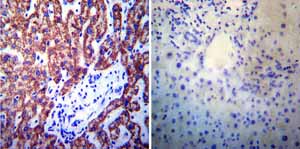 Immunohistochemistry was performed on normal biopsies of deparaffinized Human liver tissue. To expose target proteins heat induced antigen retrieval was performed using 10mM sodium citrate (pH6.0) buffer microwaved for 8-15 minutes. Following antigen retrieval tissues were blocked in 3% BSA-PBS for 30 minutes at room temperature. Tissues were then probed at a dilution of 1:200 with a mouse monoclonal antibody recognizing Presenilin 1 ab15456 or without primary antibody (negative control) overnight at 4°C in a humidified chamber. Tissues were washed extensively with PBST and endogenous peroxidase activity was quenched with a peroxidase suppressor. Detection was performed using a biotin-conjugated secondary antibody and SA-HRP followed by colorimetric detection using DAB. Tissues were counterstained with hematoxylin and prepped for mounting.
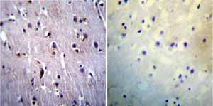 Immunohistochemistry was performed on normal biopsies of deparaffinized Human brain tissue. To expose target proteins heat induced antigen retrieval was performed using 10mM sodium citrate (pH6.0) buffer microwaved for 8-15 minutes. Following antigen retrieval tissues were blocked in 3% BSA-PBS for 30 minutes at room temperature. Tissues were then probed at a dilution of 1:20 with a mouse monoclonal antibody recognizing Presenilin 1 ab15456 or without primary antibody (negative control) overnight at 4°C in a humidified chamber. Tissues were washed extensively with PBST and endogenous peroxidase activity was quenched with a peroxidase suppressor. Detection was performed using a biotin-conjugated secondary antibody and SA-HRP followed by colorimetric detection using DAB. Tissues were counterstained with hematoxylin and prepped for mounting.
![Overlay histogram showing HepG2 cells stained with ab15456 (red line). The cells were fixed with 80% methanol (5 min) and then permeabilized with 0.1% PBS-Tween for 20 min. The cells were then incubated in 1x PBS / 10% normal goat serum / 0.3M glycine to block non-specific protein-protein interactions followed by the antibody (ab15456, 1ug/1x106 cells) for 30 min at 22ºC. The secondary antibody used was DyLight® 488 goat anti-mouse IgG1 (H+L) (ab96879) at 1/500 dilution for 30 min at 22ºC. Isotype control antibody (black line) was Mouse IgG1 [ICIGG1] (ab91353, 2µg/1x106 cells) used under the same conditions. Acquisition of >5,000 events was performed. This antibody gave a positive signal in HepG2 cells fixed with 4% paraformaldehyde (10 min) permeabilized with 0.1% PBS-Tween for 20 min used under the same conditions.](http://www.bioprodhub.com/system/product_images/ab_products/2/sub_4/15760_Presenilin-1-Primary-antibodies-ab15456-3.jpg) Overlay histogram showing HepG2 cells stained with ab15456 (red line). The cells were fixed with 80% methanol (5 min) and then permeabilized with 0.1% PBS-Tween for 20 min. The cells were then incubated in 1x PBS / 10% normal goat serum / 0.3M glycine to block non-specific protein-protein interactions followed by the antibody (ab15456, 1ug/1x106 cells) for 30 min at 22ºC. The secondary antibody used was DyLight® 488 goat anti-mouse IgG1 (H+L) (ab96879) at 1/500 dilution for 30 min at 22ºC. Isotype control antibody (black line) was Mouse IgG1 [ICIGG1] (ab91353, 2µg/1x106 cells) used under the same conditions. Acquisition of >5,000 events was performed. This antibody gave a positive signal in HepG2 cells fixed with 4% paraformaldehyde (10 min) permeabilized with 0.1% PBS-Tween for 20 min used under the same conditions.
Overlay histogram showing HepG2 cells stained with ab15456 (red line). The cells were fixed with 80% methanol (5 min) and then permeabilized with 0.1% PBS-Tween for 20 min. The cells were then incubated in 1x PBS / 10% normal goat serum / 0.3M glycine to block non-specific protein-protein interactions followed by the antibody (ab15456, 1ug/1x106 cells) for 30 min at 22ºC. The secondary antibody used was DyLight® 488 goat anti-mouse IgG1 (H+L) (ab96879) at 1/500 dilution for 30 min at 22ºC. Isotype control antibody (black line) was Mouse IgG1 [ICIGG1] (ab91353, 2µg/1x106 cells) used under the same conditions. Acquisition of >5,000 events was performed. This antibody gave a positive signal in HepG2 cells fixed with 4% paraformaldehyde (10 min) permeabilized with 0.1% PBS-Tween for 20 min used under the same conditions.
Product References
An interactive network of elastase, secretases, and PAR-2 protein regulates CXCR1 - An interactive network of elastase, secretases, and PAR-2 protein regulates CXCR1
Bakele M, Lotz-Havla AS, Jakowetz A, Carevic M, Marcos V, Muntau AC, Gersting SW, Hartl D. J Biol Chem. 2014 Jul 25;289(30):20516-25.
Cell type-specific subcellular localization of phospho-TBK1 in response to - Cell type-specific subcellular localization of phospho-TBK1 in response to
Suzuki T, Oshiumi H, Miyashita M, Aly HH, Matsumoto M, Seya T. PLoS One. 2013 Dec 9;8(12):e83639.
Interactome mapping suggests new mechanistic details underlying Alzheimer's - Interactome mapping suggests new mechanistic details underlying Alzheimer's
Soler-Lopez M, Zanzoni A, Lluis R, Stelzl U, Aloy P. Genome Res. 2011 Mar;21(3):364-76.
HPRT deficiency coordinately dysregulates canonical Wnt and presenilin-1 - HPRT deficiency coordinately dysregulates canonical Wnt and presenilin-1
Kang TH, Guibinga GH, Jinnah HA, Friedmann T. PLoS One. 2011 Jan 28;6(1):e16572.
Analysis of oestrogen regulation of alpha-, beta- and gamma-secretase gene and - Analysis of oestrogen regulation of alpha-, beta- and gamma-secretase gene and
Nord LC, Sundqvist J, Andersson E, Fried G. Neurodegener Dis. 2010;7(6):349-64.
Simvastatin increases notch signaling activity and promotes arteriogenesis after - Simvastatin increases notch signaling activity and promotes arteriogenesis after
Zacharek A, Chen J, Cui X, Yang Y, Chopp M. Stroke. 2009 Jan;40(1):254-60.