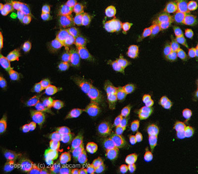
ab3765 stained HCT116 cells. The cells were 100% methanol fixed for 5 minutes at room temperature and then incubated in 1%BSA / 10% normal goat serum / 0.3M glycine in 0.1% PBS-Tween for 1hour at room temperature to permeabilise the cells and block non-specific protein-protein interactions. The cells were then incubated with the antibody (ab3765 at 10µg/ml) overnight at +4°C. The secondary antibody (pseudo-colored green) was 150081 used at a 1/1000 dilution for 1hour at room temperature. Alexa Fluor® 594 WGA was used to label plasma membranes (pseudo-colored red) at a 1/200 dilution for 1hour at room temperature. DAPI was used to stain the cell nuclei (pseudo-colored blue) at a concentration of 1.43µM for 1hour at room temperature.


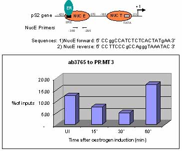
Sonicated Chromatin prepared from untreated or 17beta-estradiol (E2) treated MCF7 cells was subjected to the ChIP procedure with ab3765 to PRMT3 and the immunoprecipitated chromatin was analysed in the proximal region of the estrogen-responsive pS2 promoter (as shown above) and quantified by real-time PCR (values are % of inputs). The primers are designed to follow the nucleosome E (including the Estrogen Responsive Element ERE). 10 µl of ab3765 and 2x10⁶ cells were used in each ChIP experiment.

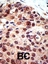
ab3765 staining PRMT3 in human breast carcinoma (BC) tissue by Immunohistochemistry (Formalin/PFA-fixed paraffin-embedded sections).


