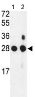
All lanes : Anti-Proteasome beta 1 antibody - C-terminal (ab135830) at 1/100 dilutionLane 1 : Mouse NIH 3T3 cell lysateLane 2 : Mouse bladder tissue lysateLysates/proteins at 35 µg per lane.
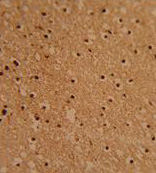
Immunohistochemical analysis of Formalin-fixed, paraffin-embedded Human brain tissue labelling Proteasome beta 1 with ab135830 at 1/50 followed by peroxidase-conjugated secondary antibody and DAB staining.
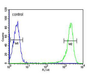
Flow cytometric analysis of HL60 cells labelling Proteasome beta 1 with ab135830 at 1/10 dilution (green) compared to a negative control cell (blue). FITC-conjugated goat-anti-rabbit secondary antibodies were used for the analysis.
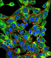
Confocal Immunofluorescent analysis of HepG2 cells labelling Proteasome beta 1 with ab135830 at 1/10 dilution followed by Alexa Fluor® 488-conjugated goat anti-rabbit lgG (green). Actin filaments have been labeled with Alexa Fluor 555 phalloidin (red). DAPI was used to stain the cell nuclear (blue).