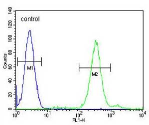
Flow cytometric analysis of HL60 cells labeling Protein cornichon homolog 2 with ab170290 at 1/10 dilution (right histogram) compared to a negative control cell (left histogram) . FITC-conjugated goat-anti-rabbit secondary antibodies were used for the analysis.
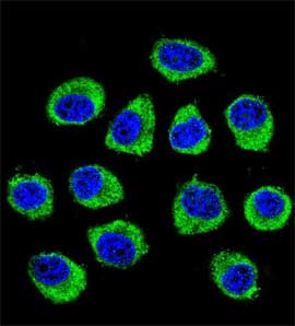
Confocal immunofluorescent analysis of U251MG cells labeling Protein cornichon homolog 2 with ab170290 at 1/10 dilution followed by Alexa Fluor 488-conjugated goat anti-rabbit lgG (green). DAPI was used to stain the cell nuclear (blue).
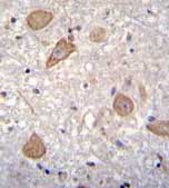
Immunohistochemical analysis of formalin-fixed, paraffin-embedded Human brain tissue labeling Protein cornichon homolog 2 with ab170290 at 1/10 dilution followed by peroxidase-conjugated secondary antibody and DAB staining.
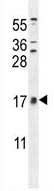
Anti-Protein cornichon homolog 2 antibody - N-terminal (ab170290) at 1/100 dilution + HL60 cell line lysate at 35 µg