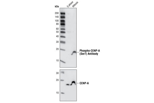
Western blot analysis of extracts from HeLa cells arrested in S phase or mitosis using Phospho-CENP-A (Ser7) Antibody (upper panel) or CENP-A Antibody #2186 (lower panel). S phase cells were treated for 12 hours with thymidine (2 mM), rinsed three times, released into normal growth medium for 10 hours and then treated an additional 12 hours with thymidine before harvesting. Mitotic cells were treated for 12 hours with thymidine, rinsed three times and then treated for 16 hours with paxitaxol (500 nM final).

Confocal immunofluorescent analysis of a mitotic HeLa cell using Phospho-CENP-A (Ser7) Antibody (green fluorescence, appearing as white in the composite image) and β-Tubulin (9F3) Rabbit mAb (Alexa Fluor® 555 Conjugate) #2116 (red). Phospho-CENP-A signal is localized to bright spots in the metaphase plate. Blue pseudocolor = DRAQ5® #4084 (fluorescent DNA dye).

