Anti-RPA70 antibody [EPR3472]
| Name | Anti-RPA70 antibody [EPR3472] |
|---|---|
| Supplier | Abcam |
| Catalog | ab79398 |
| Prices | $99.00, $397.00 |
| Sizes | 10 µl, 100 µl |
| Host | Rabbit |
| Clonality | Monoclonal |
| Isotype | IgG |
| Clone | EPR3472 |
| Applications | WB IP IHC-P ICC/IF FC Radioimmunoprecipitation |
| Species Reactivities | Human |
| Antigen | A synthetic peptide corresponding to residues in human RPA70 |
| Description | Rabbit Monoclonal |
| Gene | RPA1 |
| Conjugate | Unconjugated |
| Supplier Page | Shop |
Product images
![All lanes : Anti-RPA70 antibody [EPR3472] (ab79398) at 1/5000 dilutionLane 1 : A549 cell lysateLane 2 : HeLa cell lysateLysates/proteins at 10 µg per lane.SecondaryGoat anti-rabbit HRP at 1/2000 dilution](http://www.bioprodhub.com/system/product_images/ab_products/2/sub_4/24527_RPA70-Primary-antibodies-ab79398-1.jpg) All lanes : Anti-RPA70 antibody [EPR3472] (ab79398) at 1/5000 dilutionLane 1 : A549 cell lysateLane 2 : HeLa cell lysateLysates/proteins at 10 µg per lane.SecondaryGoat anti-rabbit HRP at 1/2000 dilution
All lanes : Anti-RPA70 antibody [EPR3472] (ab79398) at 1/5000 dilutionLane 1 : A549 cell lysateLane 2 : HeLa cell lysateLysates/proteins at 10 µg per lane.SecondaryGoat anti-rabbit HRP at 1/2000 dilution
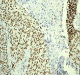 ab79398 at 1/100 dilution staining RPA70 in human cervical squamous cell carcinoma by Immunohistochemistry using paraffin-embedded tissue.
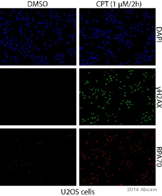 ab79398 staining RPA70 in U2OS cells by ICC/IF (Immunocytochemistry/immunofluorescence). Cells were fixed with paraformaldehyde, permeabilized with 0.5% Triton X-100 in PBS and blocked with 2% BSA for 1 hour at 25°C. Samples were incubated with primary antibody (1/500 in PBS + 0.5% Tween-20) for 2 hours at 25°C. A Cy3®-conjugated goat anti-rabbit IgG monoclonal (1/250) was used as the secondary antibody.See Abreview
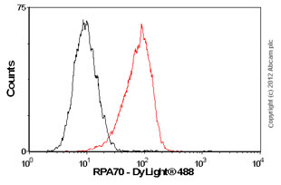 Overlay histogram showing HeLa cells stained with ab79398 (red line). The cells were fixed with 80% methanol (5 min) and then permeabilized with 0.1% PBS-Tween for 20 min. The cells were then incubated in 1x PBS / 10% normal goat serum / 0.3M glycine to block non-specific protein-protein interactions followed by the antibody (ab79398, 1/100 dilution) for 30 min at 22ºC. The secondary antibody used was DyLight® 488 goat anti-rabbit IgG (H+L) (ab96899) at 1/500 dilution for 30 min at 22ºC. Isotype control antibody (black line) was rabbit IgG (monoclonal) (1µg/1x106 cells) used under the same conditions. Acquisition of >5,000 events was performed.
Product References
ATR prohibits replication catastrophe by preventing global exhaustion of RPA. - ATR prohibits replication catastrophe by preventing global exhaustion of RPA.
Toledo LI, Altmeyer M, Rask MB, Lukas C, Larsen DH, Povlsen LK, Bekker-Jensen S, Mailand N, Bartek J, Lukas J. Cell. 2013 Nov 21;155(5):1088-103.
Kaposi's sarcoma-associated herpesvirus noncoding polyadenylated nuclear RNA - Kaposi's sarcoma-associated herpesvirus noncoding polyadenylated nuclear RNA
Rossetto CC, Pari GS. J Virol. 2011 Dec;85(24):13290-7.