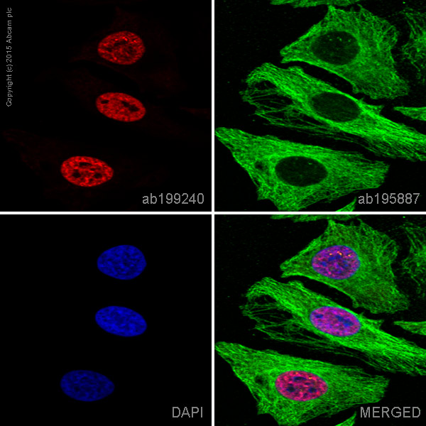
ab199240 staining RPA70 in HeLa cells. The cells were fixed with 100% methanol (5 min), permeabilized with 0.1% Triton X-100 for 5 minutes and then blocked with 1% BSA/10% normal goat serum/0.3M glycine in 0.1% PBS-Tween for 1h. The cells were then incubated overnight at +4°C with ab199240 at a 1/100 dilution (shown in red) and ab195887, Mouse monoclonal to alpha Tubulin (Alexa Fluor® 488), at a 1/250 dilution (shown in green). Nuclear DNA was labelled with DAPI (shown in blue).Image was taken with a confocal microscope (Leica-Microsystems, TCS SP8).This product also gave a positive signal under the same testing conditions in HeLa cells fixed with 4% formaldehyde (10 min).
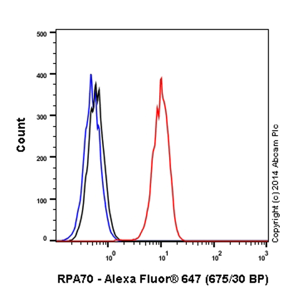
Overlay histogram showing HeLa cells stained with ab199240 (red line). The cells were fixed with 80% methanol (5 min) and then permeabilized with 0.1% PBS-Tween for 20 min. The cells were then incubated in 1x PBS / 10% normal goat serum / 0.3M glycine to block non-specific protein-protein interactions followed by the antibody (ab199240, 1/50 dilution) for 30 min at 22°C. Isotype control antibody (black line) was rabbit IgG (monoclonal) Alexa Fluor® 647 (ab199093) used at the same concentration and conditions as the primary antibody. Unlabelled sample (blue line) was also used as a control. Acquisition of >5,000 events were collected using a solid-state 25mW red diode laser (635 nm) and 675/30 bandpass filter. This antibody gave a positive signal in HeLa cells fixed with 4% formaldehyde (10 min)/permeabilized with 0.1% PBS-Tween for 20 min used under the same conditions.