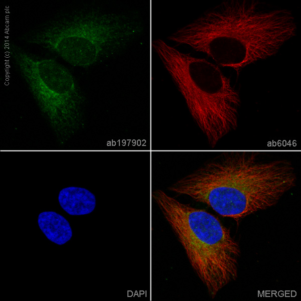
ab197902 staining SDHB in HeLa cells. The cells were fixed with 4% formaldehyde (10min), permeabilized with 0.1% Triton X-100 for 5 minutes and then blocked with 1% BSA/10% normal goat serum/0.3M glycine in 0.1% PBS-Tween for 1h. The cells were then incubated overnight at +4°C with ab197902 at 1/250 dilution (shown in green) and ab6046 (Rabbit polyclonal to beta Tubulin) at 1µg/ml. This was followed by an incubation at room temperature for 1h with ab150088, Goat polyclonal Secondary Antibody to Rabbit IgG - H&L (Alexa Fluor® 594), pre-adsorbed, at 1µg/ml (shown in red). Nuclear DNA was labelled with DAPI (shown in blue).Image was taken with a confocal microscope (Leica-Microsystems, TCS SP8).