Anti-Serum Amyloid A antibody [115]
| Name | Anti-Serum Amyloid A antibody [115] |
|---|---|
| Supplier | Abcam |
| Catalog | ab687 |
| Prices | $387.00 |
| Sizes | 100 µg |
| Host | Mouse |
| Clonality | Monoclonal |
| Isotype | IgG1 |
| Clone | 115 |
| Applications | WB IHC-P ELISA |
| Species Reactivities | Human |
| Antigen | Highly purified recombinant human serum amyloid A (MW: 12 kDa) |
| Description | Mouse Monoclonal |
| Gene | SAA1 |
| Conjugate | Unconjugated |
| Supplier Page | Shop |
Product images
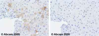 Ab687 staining human normal liver. Staining is localised to the cytoplasm.Left panel: with primary antibody at 2 ug/ml. Right panel: isotype control.Sections were stained using an automated system DAKO Autostainer Plus , at room temperature. Sections were rehydrated and antigen retrieved with the Dako 3-in-1 AR buffer EDTA pH 9.0 in a DAKO PT Link. Slides were peroxidase blocked in 3% H2O2 in methanol for 10 minutes. They were then blocked with Dako Protein block for 10 minutes (containing casein 0.25% in PBS), then incubated with primary antibody for 20 minutes, and detected with Dako Envision Flex amplification kit for 30 minutes. Colorimetric detection was completed with diaminobenzidine for 5 minutes. Slides were counterstained with Haematoxylin and coverslipped under DePeX. Please note that for manual staining we recommend to optimize the primary antibody concentration and incubation time (overnight incubation), and amplification may be required.
![All lanes : Anti-Serum Amyloid A antibody [115] (ab687) at 1/4000 dilutionLane 1 : Acute phase plasma samplesLane 2 : Acute phase plasma samplesLane 3 : Acute phase plasma samplesLane 4 : Acute phase plasma samplesLane 5 : Recombinant C-SAALane 6 : Recombinant A-SAALysates/proteins at 14 µg per lane.SecondaryGoat anti-mouse IgG-HRP at 1/10000 dilutiondeveloped using the ECL techniqueExposure time : 5 secondsImage courtesy of an anonymous Abreview.](http://www.bioprodhub.com/system/product_images/ab_products/2/sub_4/28772_Serum-Amyloid-A-Primary-antibodies-ab687-14.jpg) All lanes : Anti-Serum Amyloid A antibody [115] (ab687) at 1/4000 dilutionLane 1 : Acute phase plasma samplesLane 2 : Acute phase plasma samplesLane 3 : Acute phase plasma samplesLane 4 : Acute phase plasma samplesLane 5 : Recombinant C-SAALane 6 : Recombinant A-SAALysates/proteins at 14 µg per lane.SecondaryGoat anti-mouse IgG-HRP at 1/10000 dilutiondeveloped using the ECL techniqueExposure time : 5 secondsImage courtesy of an anonymous Abreview.
All lanes : Anti-Serum Amyloid A antibody [115] (ab687) at 1/4000 dilutionLane 1 : Acute phase plasma samplesLane 2 : Acute phase plasma samplesLane 3 : Acute phase plasma samplesLane 4 : Acute phase plasma samplesLane 5 : Recombinant C-SAALane 6 : Recombinant A-SAALysates/proteins at 14 µg per lane.SecondaryGoat anti-mouse IgG-HRP at 1/10000 dilutiondeveloped using the ECL techniqueExposure time : 5 secondsImage courtesy of an anonymous Abreview.
![Anti-Serum Amyloid A antibody [115] (ab687) at 0.5 µg/ml + Active human Serum Amyloid A full length protein (ab50232) at 0.01 µgSecondaryGoat Anti-Mouse IgG H&L (HRP) preadsorbed (ab97040) at 1/5000 dilutiondeveloped using the ECL techniquePerformed under reducing conditions.Exposure time : 1 minute](http://www.bioprodhub.com/system/product_images/ab_products/2/sub_4/28773_Serum-Amyloid-A-Primary-antibodies-ab687-18.jpg) Anti-Serum Amyloid A antibody [115] (ab687) at 0.5 µg/ml + Active human Serum Amyloid A full length protein (ab50232) at 0.01 µgSecondaryGoat Anti-Mouse IgG H&L (HRP) preadsorbed (ab97040) at 1/5000 dilutiondeveloped using the ECL techniquePerformed under reducing conditions.Exposure time : 1 minute
Anti-Serum Amyloid A antibody [115] (ab687) at 0.5 µg/ml + Active human Serum Amyloid A full length protein (ab50232) at 0.01 µgSecondaryGoat Anti-Mouse IgG H&L (HRP) preadsorbed (ab97040) at 1/5000 dilutiondeveloped using the ECL techniquePerformed under reducing conditions.Exposure time : 1 minute
Product References
Serum amyloid A inhibits apoptosis of human neutrophils via a P2X7-sensitive - Serum amyloid A inhibits apoptosis of human neutrophils via a P2X7-sensitive
Christenson K, Bjorkman L, Tangemo C, Bylund J. J Leukoc Biol. 2008 Jan;83(1):139-48. Epub 2007 Oct 25.
Inflammatory protein profile during systemic high dose interleukin-2 - Inflammatory protein profile during systemic high dose interleukin-2
Rossi L, Martin BM, Hortin GL, White RL, Foster M, Moharram R, Stroncek D, Wang E, Marincola FM, Panelli MC. Proteomics. 2006 Jan;6(2):709-20.