![Anti-SGSH antibody [EPR17312] (ab200346) at 1/1000 dilution + HepG2 (Human liver hepatocellular carcinoma) whole cell lysate at 20 µgSecondaryGoat Anti-Rabbit IgG, (H+L), Peroxidase conjugated at 1/1000 dilution](http://www.bioprodhub.com/system/product_images/ab_products/2/sub_4/29351_ab200346-242782-ab200346--wb-1.jpg)
Anti-SGSH antibody [EPR17312] (ab200346) at 1/1000 dilution + HepG2 (Human liver hepatocellular carcinoma) whole cell lysate at 20 µgSecondaryGoat Anti-Rabbit IgG, (H+L), Peroxidase conjugated at 1/1000 dilution
![Anti-SGSH antibody [EPR17312] (ab200346) at 1/10000 dilution + MCF7 (Human breast adenocarcinoma cell line) whole cell lysate at 10 µgSecondaryGoat Anti-Rabbit IgG, (H+L), Peroxidase conjugated at 1/1000 dilution](http://www.bioprodhub.com/system/product_images/ab_products/2/sub_4/29352_ab200346-242781-ab200346--wb-2.jpg)
Anti-SGSH antibody [EPR17312] (ab200346) at 1/10000 dilution + MCF7 (Human breast adenocarcinoma cell line) whole cell lysate at 10 µgSecondaryGoat Anti-Rabbit IgG, (H+L), Peroxidase conjugated at 1/1000 dilution
![Anti-SGSH antibody [EPR17312] (ab200346) at 1/10000 dilution + Human prostate cancer lysate at 10 µgSecondaryAnti-Rabbit IgG (HRP), specific to the non-reduced form of IgG at 1/1000 dilution](http://www.bioprodhub.com/system/product_images/ab_products/2/sub_4/29353_ab200346-242780-ab200346--wb-3.jpg)
Anti-SGSH antibody [EPR17312] (ab200346) at 1/10000 dilution + Human prostate cancer lysate at 10 µgSecondaryAnti-Rabbit IgG (HRP), specific to the non-reduced form of IgG at 1/1000 dilution
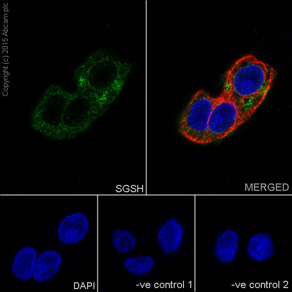
Immunofluorescent analysis of 100% methanol-fixed, 0.1% Triton X-100 permeabilized MCF7 (Human breast adenocarcinoma cell line) cells labeling SGSH with ab200346 at 1/250 dilution, followed by Goat anti-rabbit IgG (Alexa Fluor® 488) (ab150077) secondary antibody at 1/500 dilution (green). Cytoplasm staining on MCF7 cell line was observed. The nuclear counter stain is DAPI (blue). Tubulin is detected with ab7291 (anti-Tubulin mouse mAb) at 1/1000 dilution and ab150120 (AlexaFluor®594 Goat anti-Mouse secondary) at 1/500 dilution (red).The negative controls are as follows:-ve control 1: ab200346 at 1/250 dilution followed by ab150120 (AlexaFluor®594 Goat anti-Mouse secondary) at 1/500 dilution.-ve control 2: ab7291 (anti-Tubulin mouse mAb) at 1/1000 dilution followed by ab150077 (Alexa Fluor®488 Goat Anti-Rabbit IgG H&L) at 1/500 dilution.
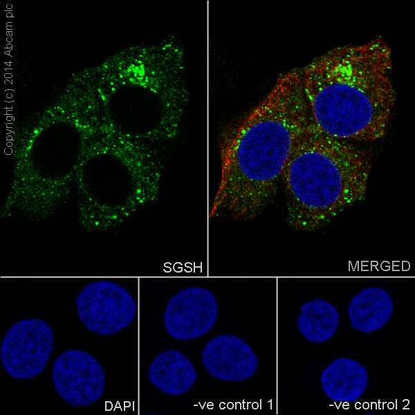
Immunofluorescent analysis of 100% methanol-fixed, 0.1% Triton X-100 permeabilized HepG2 (Human liver hepatocellular carcinoma) cells labeling SGSH with ab200346 at 1/250 dilution, followed by Goat anti-rabbit IgG (Alexa Fluor® 488) (ab150077) secondary antibody at 1/500 dilution (green). Cytoplasm staining on HepG2 cell line was observed. The nuclear counter stain is DAPI (blue). Tubulin is detected with ab7291 (anti-Tubulin mouse mAb) at 1/1000 dilution and ab150120 (AlexaFluor®594 Goat anti-Mouse secondary) at 1/500 dilution (red).The negative controls are as follows:-ve control 1: ab200346 at 1/250 dilution followed by ab150120 (AlexaFluor®594 Goat anti-Mouse secondary) at 1/500 dilution.-ve control 2: ab7291 (anti-Tubulin mouse mAb) at 1/1000 dilution followed by ab150077 (Alexa Fluor®488 Goat Anti-Rabbit IgG H&L) at 1/500 dilution.