![All lanes : Anti-SNF5 antibody [EPR12014-77] (ab192864) at 1/50000 dilutionLane 1 : 293 cell lysateLane 2 : HeLa cell lysateLane 3 : Jurkat cell lysateLane 4 : K562 cell lysateLysates/proteins at 20 µg per lane.SecondaryGoat Anti-Rabbit IgG, (H+L), Peroxidase conjugated at 1/1000 dilution](http://www.bioprodhub.com/system/product_images/ab_products/2/sub_5/2756_ab192864-230653-ab1928641.jpg)
All lanes : Anti-SNF5 antibody [EPR12014-77] (ab192864) at 1/50000 dilutionLane 1 : 293 cell lysateLane 2 : HeLa cell lysateLane 3 : Jurkat cell lysateLane 4 : K562 cell lysateLysates/proteins at 20 µg per lane.SecondaryGoat Anti-Rabbit IgG, (H+L), Peroxidase conjugated at 1/1000 dilution

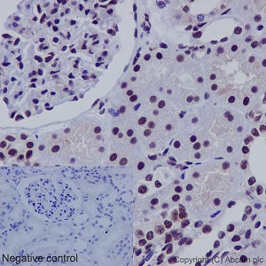
Immunohistochemical analysis of paraffin embedded Human kidney tissue sections labeling SNF5 using ab192864 at a 1/1000 dilution. A prediluted HRP Polymer for Rabbit IgG was used as the secondary. Hematoxylin counterstain.

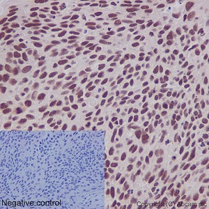
Immunohistochemical analysis of paraffin embedded Human squamous cell carcinoma of cervix tissue sections labeling SNF5 using ab192864 at a 1/1000 dilution. A predilutedHRP Polymer for Rabbit IgG was used as the secondary. Hematoxylin counterstain.

Immunofluorescent analysis of 4% paraformaldehyde fixed HeLa cells labeling SNF5 using ab192864 at a 1/500 dilution. A Goat anti rabbit IgG (Alexa Fluor®488) (ab150077) was used as the secondary at a 1/400 dilution. Counterstain DAPI. Cells were permeabilized using 0.1% Triton X-100.

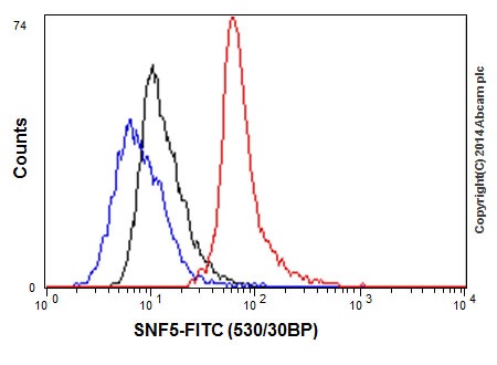
Flow cytometric analysis of Jurkat cells labeling SNF5 using ab192864 at a 1/110 dilution (red). Goat anti rabbit IgG (FITC) used as the secondary antibody at a 1/150 dilution. Isotype control Rabbit monoclonal IgG (black). Unlabeled/control cells without incubation with primary and secondary antibody (blue). Cells were fixed in 2% paraformaldehyde.

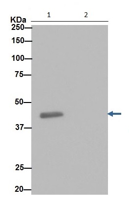
Western blot analysis of K562 cell lysate immunoprecipitated using ab192864 at a 1/30 dilution (lane 1).Lane 2: PBS instead of K562 lysateSecondary antibody was anti-rabbit IgG (HRP) specific to the non-reduced form of IgG at a 1/1500 dilution.
![All lanes : Anti-SNF5 antibody [EPR12014-77] (ab192864) at 1/50000 dilutionLane 1 : 293 cell lysateLane 2 : HeLa cell lysateLane 3 : Jurkat cell lysateLane 4 : K562 cell lysateLysates/proteins at 20 µg per lane.SecondaryGoat Anti-Rabbit IgG, (H+L), Peroxidase conjugated at 1/1000 dilution](http://www.bioprodhub.com/system/product_images/ab_products/2/sub_5/2756_ab192864-230653-ab1928641.jpg)


