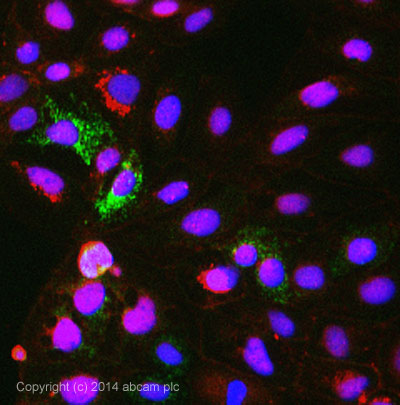
ICC/IF image of ab71978 stained CACO-2 cells. The cells were 4% formaldehyde fixed (10 min) and then incubated in 1%BSA / 10% normal goat serum / 0.3M glycine in 0.1% PBS-Tween for 1h to permeabilise the cells and block non-specific protein-protein interactions. The cells were then incubated with the antibody ab71978 at 5µg/ml overnight at +4°C. The secondary antibody (pseudo-colored green) was Alexa Fluor® 488 goat anti- mouse (ab150117) IgG (H+L) preadsorbed, used at a 1/1000 dilution for 1h. Alexa Fluor® 594 WGA was used to label plasma membranes (pseudo-colored red) at a 1/200 dilution for 1h at room temperature. DAPI was used to stain the cell nuclei (pseudo-colored blue) at a concentration of 1.43µM for 1hour at room temperature.
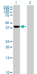 All lanes : Anti-SQRDL antibody (ab71978) at 1/500 dilutionLane 1 : SQRDL transfected 293T cell lysateLane 2 : Non transfected 293T cell lysateLysates/proteins at 25 µg per lane.SecondaryGoat anti-mouse IgG (H&L)-HRP conjugate at 1/2500 dilution