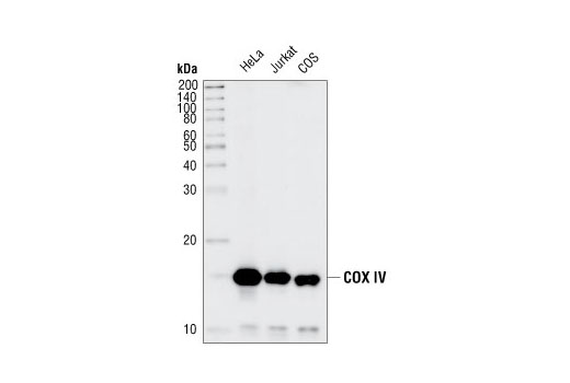
Western blot analysis of extracts from HeLa, Jurkat and COS cell lines, using COX IV (3E11) Rabbit mAb.

Immunohistochemical analysis of paraffin-embedded human colon carcinoma, showing staining of the mitochondria, using COX IV (3E11) Rabbit mAb.

Immunohistochemical analysis of paraffin-embedded human breast carcinoma, using COX IV (3E11) Rabbit mAb in the presence of control peptide (left) or Cox IV Blocking Peptide #1034 (right).

Immunohistochemical analysis of paraffin-embedded H1650 xenograft, using COX IV Rabbit mAb. Note specific staining of human cancer cells.

Immunohistochemical analysis of frozen H1650 xenograft, using Cox IV (3E11) Rabbit mAb.

Confocal immunofluorescent analysis of HeLa cells labeled with COX IV (3E11) Rabbit mAb (green). Actin filaments have been labeled with Alexa Fluor® 555 phalloidin (red). Blue pseudocolor = DRAQ5 ® #4084 (fluorescent DNA dye).

Flow cytometric analysis of HeLa cells, using COX IV (3E11) Rabbit mAb (blue) compared to a nonspecific negative control antibody (red).