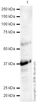
Anti-Synaptophysin antibody (ab23754) at 1 µg/ml + Human brain tissue lysate at 20 µgSecondaryGoat polyclonal to Rabbit IgG (Alexa Fluor® 680) at 1/10000 dilutionPerformed under reducing conditions.
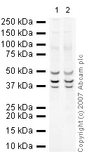
All lanes : Anti-Synaptophysin antibody (ab23754) at 1 µg/mlLane 1 : Brain (Mouse) Tissue Lysate - normal tissueLane 2 : Brain (Rat) Tissue Lysate - normal tissue Lysates/proteins at 10 µg per lane.SecondaryIRDye 680 Conjugated Goat Anti-Rabbit IgG (H+L) at 1/10000 dilutionPerformed under reducing conditions.
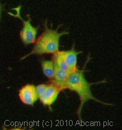
ICC/IF image of ab23754 stained PC12 cells. The cells were 100% methanol fixed (5 min) and then incubated in 1%BSA / 10% normal goat serum / 0.3M glycine in 0.1% PBS-Tween for 1h to permeabilise the cells and block non-specific protein-protein interactions. The cells were then incubated with the antibody (ab23754, 5µg/ml) overnight at +4°C. The secondary antibody (green) was Alexa Fluor® 488 goat anti-rabbit IgG (H+L) used at a 1/1000 dilution for 1h. Alexa Fluor® 594 WGA was used to label plasma membranes (red) at a 1/200 dilution for 1h. DAPI was used to stain the cell nuclei (blue) at a concentration of 1.43µM.
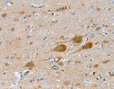
IHC image of Synaptophysin staining in human cerebral cortex FFPE section, performed on a BondTM system using the standard protocol F. The section was pre-treated using heat mediated antigen retrieval with sodium citrate buffer (pH6, epitope retrieval solution 1) for 20 mins. The section was then incubated with ab23754, 1µg/ml, for 15 mins at room temperature and detected using an HRP conjugated compact polymer system. DAB was used as the chromogen. The section was then counterstained with haematoxylin and mounted with DPX.