
All lanes : Anti-TCEB2 antibody (ab45297) at 1/1000 dilutionLane 1 : Recombinant TCEB2 protein (15ug/lane)Lane 2 : Negative control protein

All lanes : Anti-TCEB2 antibody (ab45297) at 1 µg/mlLane 1 : HeLa (Human epithelial carcinoma cell line) Whole Cell Lysate (ab27252) at 20 µgLane 2 : HeLa (Human epithelial carcinoma cell line) Nuclear Lysate (ab27251) at 20 µgLane 3 : Skeletal Muscle Normal (Human) Tissue Lysate (ab15370) at 20 µgLane 4 : Negative control - Small Intestine (Human) Tissue Lysate (ab7923)at 20 µg SecondaryRabbit IgG secondary antibody (ab28446) at 1/5000 dilution
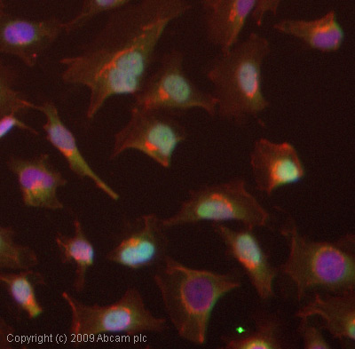
ICC/IF image of ab45297 stained HeLa cells. The cells were 4% PFA fixed (10 min) and then incubated in 1%BSA / 10% normal goat serum / 0.3M glycine in 0.1% PBS-Tween for 1h to permeabilise the cells and block non-specific protein-protein interactions. The cells were then incubated with the antibody (ab45297, 1µg/ml) overnight at +4°C. The secondary antibody (green) was Alexa Fluor® 488 goat anti-rabbit IgG (H+L) used at a 1/1000 dilution for 1h. Alexa Fluor® 594 WGA was used to label plasma membranes (red) at a 1/200 dilution for 1h. DAPI was used to stain the cell nuclei (blue) at a concentration of 1.43µM.