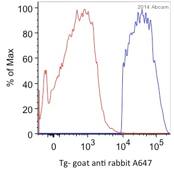
ab156008 staining Thyroglobulin in mouse thyroid cells by Flow Cytometry. Cells were fixed with formaldehyde and permeabilized with permeabilization buffer. The sample was incubated with the primary antibody (1/100 in FACS buffer) for 30 minutes at 24°C. An Alexa Fluor® 647-conjugated goat anti-rabbit IgG (1/2000) was used as the secondary antibody. Gating Strategy: Epithlial cells. Red line shows unlabeled sample, blue line shows labeled sample.See Abreview
![All lanes : Anti-Thyroglobulin antibody [EPR9730] (ab156008) at 1/10000 dilutionLane 1 : Mouse thyroid lysateLane 2 : Rat thyroid lysateLane 3 : Human thyroid lysateLysates/proteins at 10 µg per lane.SecondaryHRP labelled goat anti-rabbit at 1/2000 dilution](http://www.bioprodhub.com/system/product_images/ab_products/2/sub_5/10888_Thyroglobulin-Primary-antibodies-ab156008-1.jpg)
All lanes : Anti-Thyroglobulin antibody [EPR9730] (ab156008) at 1/10000 dilutionLane 1 : Mouse thyroid lysateLane 2 : Rat thyroid lysateLane 3 : Human thyroid lysateLysates/proteins at 10 µg per lane.SecondaryHRP labelled goat anti-rabbit at 1/2000 dilution
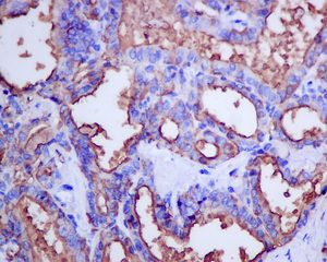
Immunohistochemical analysis of paraffin embedded Human thyroid gland follicular carcinoma tissue labeling Thyroglobulin with ab156008 antibody at 1/250.
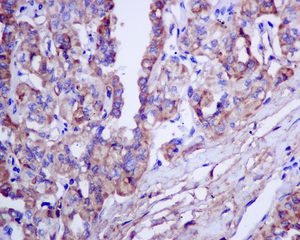
Immunohistochemical analysis of paraffin embedded Human thyroid gland papillary carcinoma tissue labeling Thyroglobulin with ab156008 antibody at 1/250.
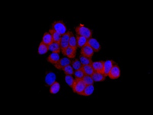
Immunofluorescent analysis of TT cells labeling Thyroglobulin with ab156008 at 1/50.