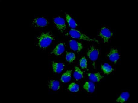
Immunofluorescence analysis of paraformaldehyde-fixed HeLa cells, labeling TOMM20 (green) with ab186734 at 1/250 dilution. Alexa Fluor®488-conjugated goat anti-rabbit IgG was used as a secondary antibody at 1/200 dilution. Nuclei were counterstained with DAPI (blue).
![Anti-TOMM20 antibody [EPR15581] - Mitochondrial Marker (ab186734) at 1/5000 dilution + HepG2 cell lysate at 20 µgSecondaryGoat Anti-Rabbit IgG, (H+L), Peroxidase conjugate at 1/1000 dilution](http://www.bioprodhub.com/system/product_images/ab_products/2/sub_5/13064_ab186734-219953-ab186734WB.jpg)
Anti-TOMM20 antibody [EPR15581] - Mitochondrial Marker (ab186734) at 1/5000 dilution + HepG2 cell lysate at 20 µgSecondaryGoat Anti-Rabbit IgG, (H+L), Peroxidase conjugate at 1/1000 dilution
![Anti-TOMM20 antibody [EPR15581] - Mitochondrial Marker (ab186734) at 1/20000 dilution + HeLa cell lysate at 20 µgSecondaryGoat Anti-Rabbit IgG, (H+L), Peroxidase conjugate at 1/1000 dilution](http://www.bioprodhub.com/system/product_images/ab_products/2/sub_5/13065_ab186734-219952-ab186734WBb.jpg)
Anti-TOMM20 antibody [EPR15581] - Mitochondrial Marker (ab186734) at 1/20000 dilution + HeLa cell lysate at 20 µgSecondaryGoat Anti-Rabbit IgG, (H+L), Peroxidase conjugate at 1/1000 dilution
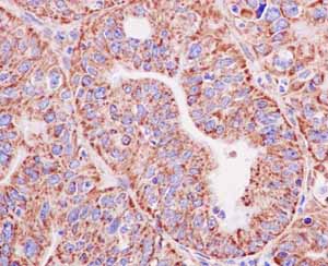
Immunohistochemical analysis of paraffin-embedded Human endometrial adenocarcinoma tissue, labeling TOMM20 with ab186734 at 1/100 dilution. Detected using HRP Polymer for Rabbit IgG and counter-stained using hematoxylin.
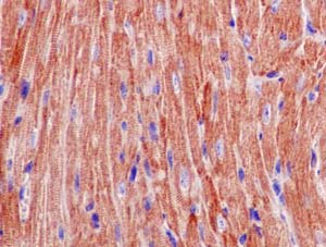
Immunohistochemical analysis of paraffin-embedded mouse cardiac muscle tissue tissue, labeling TOMM20 with ab186734 at 1/100 dilution. Detected using HRP Polymer for Rabbit IgG and counter-stained using hematoxylin.
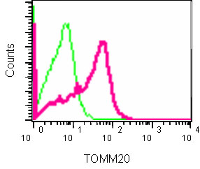
Flow cytometry analysis of TOMM20 expression in NCI-H2228 cells using ab186734 at 1/40 dilution (red) and a rabbit IgG as negative control (green).