![All lanes : Anti-TRAP 5 antibody [EPR15556] (ab191406) at 1/10000 dilutionLane 1 : A431 cell lysateLane 2 : MDA-MB435 cell lysateLysates/proteins at 20 µg per lane.SecondaryGoat anti-rabbit IgG, (H+L), peroxidase conjugated at 1/1000 dilution](http://www.bioprodhub.com/system/product_images/ab_products/2/sub_5/14116_ab191406-228744-ab191406WB1.jpg)
All lanes : Anti-TRAP 5 antibody [EPR15556] (ab191406) at 1/10000 dilutionLane 1 : A431 cell lysateLane 2 : MDA-MB435 cell lysateLysates/proteins at 20 µg per lane.SecondaryGoat anti-rabbit IgG, (H+L), peroxidase conjugated at 1/1000 dilution
![All lanes : Anti-TRAP 5 antibody [EPR15556] (ab191406) at 1/1000 dilutionLane 1 : Human tonsil lysateLane 2 : Human fetal kidney lysateLysates/proteins at 20 µg per lane.SecondaryAnti-Rabbit IgG (HRP), specific to the non-reduced form of IgG at 1/1000 dilution](http://www.bioprodhub.com/system/product_images/ab_products/2/sub_5/14117_ab191406-228743-ab191406WB2.jpg)
All lanes : Anti-TRAP 5 antibody [EPR15556] (ab191406) at 1/1000 dilutionLane 1 : Human tonsil lysateLane 2 : Human fetal kidney lysateLysates/proteins at 20 µg per lane.SecondaryAnti-Rabbit IgG (HRP), specific to the non-reduced form of IgG at 1/1000 dilution
![All lanes : Anti-TRAP 5 antibody [EPR15556] (ab191406) at 1/1000 dilutionLane 1 : C6 cell lysateLane 2 : Raw264.7 cell lysateLane 3 : PC-12 cell lysateLane 4 : NIH 3T3/cell lysateLysates/proteins at 10 µg per lane.SecondaryAnti-Rabbit IgG (HRP), specific to the non-reduced form of IgG at 1/1000 dilution](http://www.bioprodhub.com/system/product_images/ab_products/2/sub_5/14118_ab191406-228742-ab191406WB3.jpg)
All lanes : Anti-TRAP 5 antibody [EPR15556] (ab191406) at 1/1000 dilutionLane 1 : C6 cell lysateLane 2 : Raw264.7 cell lysateLane 3 : PC-12 cell lysateLane 4 : NIH 3T3/cell lysateLysates/proteins at 10 µg per lane.SecondaryAnti-Rabbit IgG (HRP), specific to the non-reduced form of IgG at 1/1000 dilution

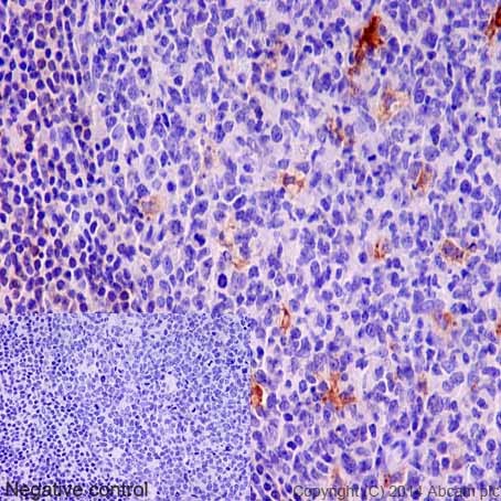
Immunohistochemical analysis of paraffin-embedded Human tonsil tissue labeling TRAP 5 with ab191406 at 1/100 dilution, followed by prediluted HRP Polymer for Rabbit/Mouse IgG. Counter stained with Hematoxylin.In the negative control PBS was used instead of primary antibody.

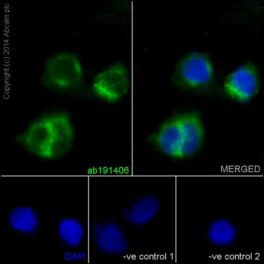
Immunofluorescent analysis of 4% paraformaldehyde-fixed, 0.1% tritonX-100 permeabilized MDA-MB435 cells labeling TRAP 5 with ab191406 at 1/100 dilution, followed by Goat anti-rabbit IgG (Alexa Fluor®488) secondary antibody (ab150077) at 1/200 dilution. Nuclear counter stain DAPI (blue).The two negative controls used anti-TRAP 5 primary antibody at 1/100 dilution, followed by Goat anti-mouse IgG (Alexa Fluor®594) secondary antibody at 1/400 dilution.

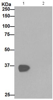
Western blot analysis of TRAP 5 in MDA-MB435 cell lysate immunoprecipitated using ab191406 at 1/50 dilution (Lane 1). Lane 2: PBS instead of MDA-MB-435 lysates.Secondary antibody: Anti-Rabbit IgG (HRP), specific to the non-reduced form of IgG at 1/1000 dilution.
![All lanes : Anti-TRAP 5 antibody [EPR15556] (ab191406) at 1/10000 dilutionLane 1 : A431 cell lysateLane 2 : MDA-MB435 cell lysateLysates/proteins at 20 µg per lane.SecondaryGoat anti-rabbit IgG, (H+L), peroxidase conjugated at 1/1000 dilution](http://www.bioprodhub.com/system/product_images/ab_products/2/sub_5/14116_ab191406-228744-ab191406WB1.jpg)
![All lanes : Anti-TRAP 5 antibody [EPR15556] (ab191406) at 1/1000 dilutionLane 1 : Human tonsil lysateLane 2 : Human fetal kidney lysateLysates/proteins at 20 µg per lane.SecondaryAnti-Rabbit IgG (HRP), specific to the non-reduced form of IgG at 1/1000 dilution](http://www.bioprodhub.com/system/product_images/ab_products/2/sub_5/14117_ab191406-228743-ab191406WB2.jpg)
![All lanes : Anti-TRAP 5 antibody [EPR15556] (ab191406) at 1/1000 dilutionLane 1 : C6 cell lysateLane 2 : Raw264.7 cell lysateLane 3 : PC-12 cell lysateLane 4 : NIH 3T3/cell lysateLysates/proteins at 10 µg per lane.SecondaryAnti-Rabbit IgG (HRP), specific to the non-reduced form of IgG at 1/1000 dilution](http://www.bioprodhub.com/system/product_images/ab_products/2/sub_5/14118_ab191406-228742-ab191406WB3.jpg)


