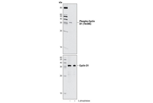
Western blot analysis of extracts from HT-1080 cells, treated with MG132 and with or without λ phosphatase, using Phospho-Cyclin D1 (Thr286) (D29B3) XP ® Rabbit mAb (upper) or Cyclin D1 (DC56) Mouse mAb #2926 (lower).

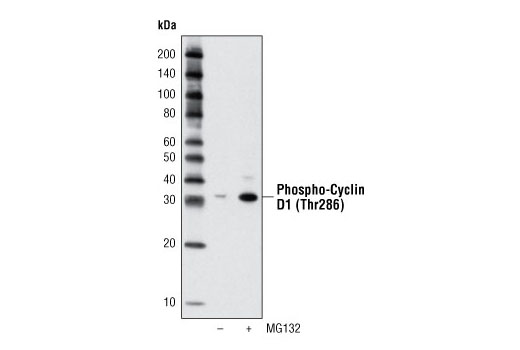
Western blot analysis of extracts from HT-1080 cells, untreated or treated with MG132 (10 μM, 4 hours), using Phospho-Cyclin D1 (Thr286) (D29B3) XP ® Rabbit mAb.

Confocal immmunofluorescent analysis of HT-1080 (top) and Saos-2 cells (bottom), untreated (left) or treated with MG132 alone (center) or with MG132 followed by λ-phosphatase (right), using Phospho-Cyclin D1 (Thr286) (D29B3) XP ® Rabbit mAb (green). Actin filaments were labeled with DY-554 phalloidin (red).

Flow cytometric analysis of HT-1080 cells, untreated (blue) or MG132-treated (green), using Phospho-Cyclin D1 (Thr286) (D29B3) XP ® Rabbit mAb.


