ab106088 at 2.5µg/ml staining TUBA1B in Formalin-fixed, Paraffin-embedded Human thyroid follicular epithelium by Immunohistochemistry. The image shows the localization of the antibody as the precipitated red signal, with a hematoxylin purple nuclear counterstain.
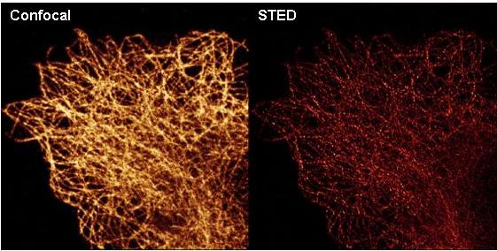
ab106088 at 0.1µg/ml staining TUBA1B in 4% paraformaldehyde fixed A459 cells by Immunofluorescence. Staining is shown using conventional confocal microscopy (left image) and by high resolution TCS STED nanoscopy (right image). DyLight488™ conjugated anti-mouse IgG secondary antibody was used for detection at 1 µg/ml.
![All lanes : Anti-TUBA1B antibody [17H11.F10] (ab106088) at 1/1000 dilution (for 1hr)Lane 1 : HeLa whole cell lysateLane 2 : HEK293 whole cell lysateLysates/proteins at 10 µg per lane.Secondaryrabbit-anti-mouse IgG HRP at 1/40000 dilution](http://www.bioprodhub.com/system/product_images/ab_products/2/sub_5/16137_TUBA1B-Primary-antibodies-ab106088-3.jpg)
All lanes : Anti-TUBA1B antibody [17H11.F10] (ab106088) at 1/1000 dilution (for 1hr)Lane 1 : HeLa whole cell lysateLane 2 : HEK293 whole cell lysateLysates/proteins at 10 µg per lane.Secondaryrabbit-anti-mouse IgG HRP at 1/40000 dilution