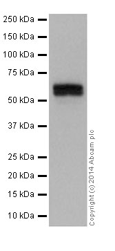
Anti-Tyrosine Hydroxylase antibody (ab191486) at 1/10000 dilution + Human fetal brain lysate at 20 µgSecondaryGoat Anti-Rabbit IgG, (H+L), Peroxidase conjugated at 1/1000 dilution
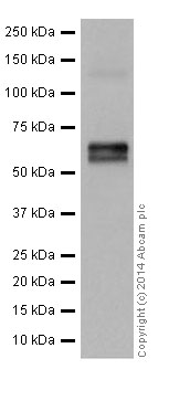
Anti-Tyrosine Hydroxylase antibody (ab191486) at 1/10000 dilution + Human adrenal gland lysate at 10 µgSecondaryAnti-Rabbit IgG (HRP), specific to the non-reduced form of IgG at 1/1000 dilution
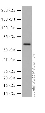
Anti-Tyrosine Hydroxylase antibody (ab191486) at 1/5000 dilution + Human cerebellum lysate at 10 µgSecondaryAnti-Rabbit IgG (HRP), specific to the non-reduced form of IgG at 1/1000 dilution
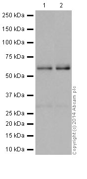
All lanes : Anti-Tyrosine Hydroxylase antibody (ab191486) at 1/2000 dilutionLane 1 : Mouse brain lysateLane 2 : Rat brain lysateLysates/proteins at 10 µg per lane.SecondaryGoat Anti-Rabbit IgG, (H+L),Peroxidase conjugated at 1/1000 dilution
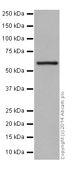
Anti-Tyrosine Hydroxylase antibody (ab191486) at 1/10000 dilution + PC-12 (Rat adrenal gland pheochromocytoma) cell lysate at 20 µgSecondaryGoat Anti-Rabbit IgG, (H+L),Peroxidase conjugated at 1/1000 dilution
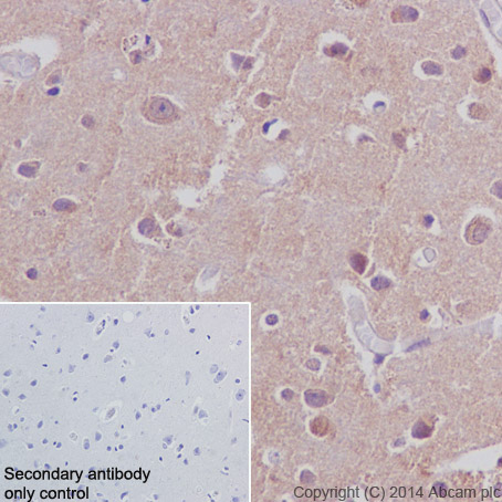
Immunohistochemical analysis of paraffin-embedded Human cerebral cortex tissue labeling Tyrosine Hydroxylase with ab191486 at 1/200 dilution, followed by Goat Anti-Rabbit IgG H&L (HRP) (ab97051) at 1/500 dilution. Nucleus and cytoplasm staining on Human cerebral cortex is observed. Counter stained with Hematoxylin.Secondary antibody only control: Using PBS instead of primary anyibody, secondary antibody is Goat Anti-Rabbit IgG H&L (HRP) (ab97051) at 1/500 dilution.
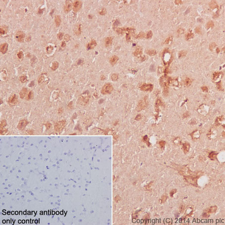
Immunohistochemical analysis of paraffin-embedded Mouse cerebral cortex tissue labeling Tyrosine Hydroxylase with ab191486 at 1/200 dilution, followed by Goat Anti-Rabbit IgG H&L (HRP) (ab97051) at 1/500 dilution. Nucleus and weakly cytoplasm staining on Mouse cerebral cortex is observed. Counter stained with Hematoxylin.Secondary antibody only control: Using PBS instead of primary anyibody, secondary antibody is Goat Anti-Rabbit IgG H&L (HRP) (ab97051) at 1/500 dilution.


