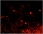
Immunohistochemical analysis of mouse dorsal root ganglia labelling VR1 with ab203103 at 10 µg/mL. Visualized with anti-mouse-Cy3 conjugate (red).
![Lane 1 : Anti-VR1 antibody [BS397] - C-terminal (ab203103) at 2 µg/ml (overnight)Lane 2 : Anti-VR1 antibody [BS397] - C-terminal (ab203103) at 2 µg/ml ((pre-absorbed))Lane 1 : Rat PC12 cell lysateLane 2 : Rat PC12 cell lysateLysates/proteins at 80 µg per lane.SecondaryAnti-mouse-HRP at 1/6000 dilutiondeveloped using the ECL technique](http://www.bioprodhub.com/system/product_images/ab_products/2/sub_5/20563_ab203103-245109-ab203103-copy.jpg)
Lane 1 : Anti-VR1 antibody [BS397] - C-terminal (ab203103) at 2 µg/ml (overnight)Lane 2 : Anti-VR1 antibody [BS397] - C-terminal (ab203103) at 2 µg/ml ((pre-absorbed))Lane 1 : Rat PC12 cell lysateLane 2 : Rat PC12 cell lysateLysates/proteins at 80 µg per lane.SecondaryAnti-mouse-HRP at 1/6000 dilutiondeveloped using the ECL technique
![All lanes : Anti-VR1 antibody [BS397] - C-terminal (ab203103) at 1 µg/mlLane 1 : controlLane 2 : Forskolin stimulated 50B11 hybrid mouse x rat DRG cell lysateLane 3 : Forskolin and NGF stimulated 50B11 hybrid mouse x rat DRG cell lysateLane 4 : NGF-stimulated PC12 cell lysate (NGF control)Lysates/proteins at 10 µg per lane.developed using the ECL technique](http://www.bioprodhub.com/system/product_images/ab_products/2/sub_5/20564_ab203103-245114-ab203103d-copy.jpg)
All lanes : Anti-VR1 antibody [BS397] - C-terminal (ab203103) at 1 µg/mlLane 1 : controlLane 2 : Forskolin stimulated 50B11 hybrid mouse x rat DRG cell lysateLane 3 : Forskolin and NGF stimulated 50B11 hybrid mouse x rat DRG cell lysateLane 4 : NGF-stimulated PC12 cell lysate (NGF control)Lysates/proteins at 10 µg per lane.developed using the ECL technique
![Lane 1 : Anti-VR1 antibody [BS397] - C-terminal (ab203103) at 2 µg/mlLane 2 : Anti-VR1 antibody [BS397] - C-terminal (ab203103) at 1 µg/mlLane 3 : Anti-VR1 antibody [BS397] - C-terminal (ab203103) at 2 µg/ml (pre-absorbed)Lane 1 : Mouse brain homogenateLane 2 : Mouse brain homogenateLane 3 : Mouse brain homogenateLysates/proteins at 25 µg per lane.SecondaryAnti-mouse-HRP at 1/6000 dilutiondeveloped using the ECL technique](http://www.bioprodhub.com/system/product_images/ab_products/2/sub_5/20565_ab203103-245113-ab203103c-copy.jpg)
Lane 1 : Anti-VR1 antibody [BS397] - C-terminal (ab203103) at 2 µg/mlLane 2 : Anti-VR1 antibody [BS397] - C-terminal (ab203103) at 1 µg/mlLane 3 : Anti-VR1 antibody [BS397] - C-terminal (ab203103) at 2 µg/ml (pre-absorbed)Lane 1 : Mouse brain homogenateLane 2 : Mouse brain homogenateLane 3 : Mouse brain homogenateLysates/proteins at 25 µg per lane.SecondaryAnti-mouse-HRP at 1/6000 dilutiondeveloped using the ECL technique

![Lane 1 : Anti-VR1 antibody [BS397] - C-terminal (ab203103) at 2 µg/ml (overnight)Lane 2 : Anti-VR1 antibody [BS397] - C-terminal (ab203103) at 2 µg/ml ((pre-absorbed))Lane 1 : Rat PC12 cell lysateLane 2 : Rat PC12 cell lysateLysates/proteins at 80 µg per lane.SecondaryAnti-mouse-HRP at 1/6000 dilutiondeveloped using the ECL technique](http://www.bioprodhub.com/system/product_images/ab_products/2/sub_5/20563_ab203103-245109-ab203103-copy.jpg)
![All lanes : Anti-VR1 antibody [BS397] - C-terminal (ab203103) at 1 µg/mlLane 1 : controlLane 2 : Forskolin stimulated 50B11 hybrid mouse x rat DRG cell lysateLane 3 : Forskolin and NGF stimulated 50B11 hybrid mouse x rat DRG cell lysateLane 4 : NGF-stimulated PC12 cell lysate (NGF control)Lysates/proteins at 10 µg per lane.developed using the ECL technique](http://www.bioprodhub.com/system/product_images/ab_products/2/sub_5/20564_ab203103-245114-ab203103d-copy.jpg)
![Lane 1 : Anti-VR1 antibody [BS397] - C-terminal (ab203103) at 2 µg/mlLane 2 : Anti-VR1 antibody [BS397] - C-terminal (ab203103) at 1 µg/mlLane 3 : Anti-VR1 antibody [BS397] - C-terminal (ab203103) at 2 µg/ml (pre-absorbed)Lane 1 : Mouse brain homogenateLane 2 : Mouse brain homogenateLane 3 : Mouse brain homogenateLysates/proteins at 25 µg per lane.SecondaryAnti-mouse-HRP at 1/6000 dilutiondeveloped using the ECL technique](http://www.bioprodhub.com/system/product_images/ab_products/2/sub_5/20565_ab203103-245113-ab203103c-copy.jpg)