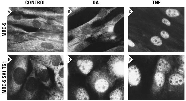
Influence of okadaic acid (OA) and tumor necrosis factor (TNF) on the translocation of NFκB p65 from the cytoplasm to the nucleus. Cells were immunostained with NFκB p65 (A): sc-109 and visualized with biotinylated goat anti-rabbit IgG and fluorescein isothiocyanate-conjugated avidin. Kindly provided by Sree Devi Menon.
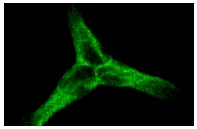
NFκB p65 (A)-G: sc-109-G. Immunofluorescence staining of methanol-fixed A-431 cells showing cytoplasmic staining.
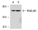
NFκB p65 (A): sc-109. Western blot analysis of NFκB p65 expression in extracts of human A-431 (A) and K-562 (B) whole cell lysates.

ChIP analysis of transcription factor binding to the Interferon β promoter before (A) and six hours after (B) Sendai virus infection of HeLa cells. Antibodies tested included NFκB p65 (A): sc-109, ATF-2 (N-96): sc-6233, GCN5 (N-18): sc-6303, Pol II (H-224): sc-9001, TFIID (TBP)(SI-1): sc-273 and CBP (A-22): sc-369. Data kindly provided by G. Mosialos.

ChIP analysis of cytokine gene promoter occupancy in TNFα-treated mouse embryonic fibroblasts. IP's permorned without (A) and with (B) primary antibody. Antibodies tested include NFκB p65 (A): sc-109, RelB (C-19): sc-226 and p300 (N-15): sc-584. Data kindly provided by A. Hoffmann.
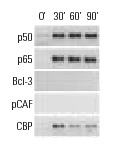
ChIP analysis of cofactor occupancy dynamics on the ICAM1 promoter in 293 cells in response to IL-1β treatment. Antibodies tested include NFκB p50 (C-19): sc-1190, NFκB p50 (E-10): sc-8414, NFκB p50 (H-119): sc-7178, NFκB p65 (C-20): sc-372, NFκB p65 (A): sc-109, NFκB p65 (H-286): sc-7151, Bcl-3 (C-14): sc-185, Bcl-3 (H-146): sc-13038, PCAF (C-16): sc-6300, PCAF (H-369): sc-8999, CBP (A-22): sc-369, CBP (C-1): sc-7300, CBP (C-20): sc-583, CBP (451): sc-1211. Data kindly provided by M.G. Rosenfeld and reproduced with permission from Baek et al., Cell 2002, 110: 55-67.
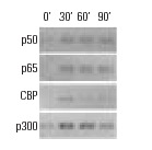
ChIP analysis of cofactor occupancy dynamics on the IL-8 promoter in 293 cells in response to IL-1β treatment. Antibodies tested include NFκB p50 (C-19): sc-1190, NFκB p50 (E-10): sc-8414, NFκB p50 (H-119): sc-7178, NFκB p65 (C-20): sc-372, NFκB p65 (A): sc-109, NFκB p65 (H-286): sc-7151, CBP (A-22): sc-369, CBP (C-1): sc-7300, CBP (C-20): sc-583, CBP (451): sc-1211, p300 (C-20): sc-sc-585, p300 (N-15): sc-584, p300 (H-272): sc-8981. Data kindly provided by M.G. Rosenfeld and reproduced with permission from Baek et al., Cell 2002, 110: 55-67.
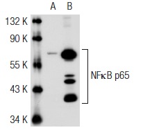
NFκB p65 (A): sc-109. Western blot analysis of NFκB p65 expression in non-transfected: sc-117752 (A) and mouse NFκB p65 transfected: sc-122027 (B) 293T whole cell lysates.
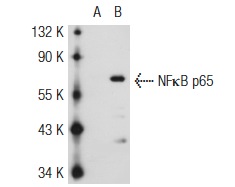
NFκB p65 (A)-G: sc-109-G. Western blot analysis of NFκB p65 expression in non-transfected: sc-117752 (A) and mouse NFκB p65 transfected: sc-122027 (B) 293T whole cell lysates.
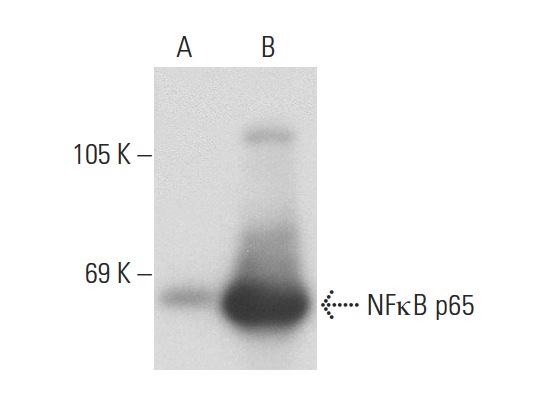
NFκB p65 (A): sc-109. Western blot analysis of NFκB p65 expression in non-transfected: sc-117752 (A) and human NFκB p65 transfected: sc-122028 (B) 293T whole cell lysates.
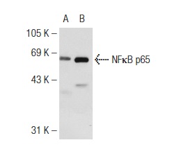
NFκB p65 (A): sc-109. Western blot analysis of NFκB p65 expression in non-transfected: sc-117752 (A) and mouse NFκB p65 transfected: sc-122027 (B) 293T whole cell lysates.
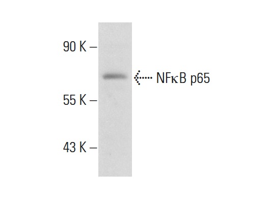
NFκB p65 (A)-G: sc-109-G. Western blot analysis of NFκB p65 expression in SK-BR-3 whole cell lysate.
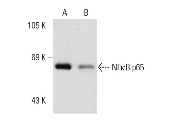
NFκB p65 (A): sc-109. Western blot analysis of NFκB p65 expression in HeLa (A) and MIA PaCa-2 (B) whole cell lysates.


