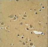
AMY2B antibody immunohistochemistry of formalin-fixed and paraffin-embedded human brain tissue followed by peroxidase-conjugated secondary antibody and DAB staining.
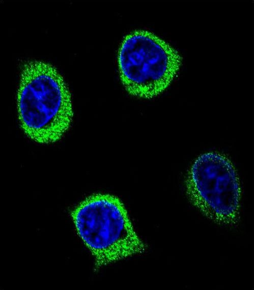
Confocal immunofluorescent of AMY2B Antibody with 293 cell followed by Alexa Fluor 488-conjugated goat anti-rabbit lgG (green). DAPI was used to stain the cell nuclear (blue).
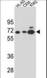
AMY2B Antibody western blot of HL-60,CEM and K562 cell line lysates (35 ug/lane). The AMY2B antibody detected the AMY2B protein (arrow).
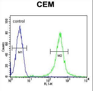
AMY2B Antibody flow cytometry of CEM cells (right histogram) compared to a negative control cell (left histogram). FITC-conjugated goat-anti-rabbit secondary antibodies were used for the analysis.