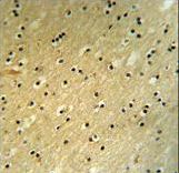
BCOR Antibody (Center S1122) IHC of formalin-fixed and paraffin-embedded mouse brain tissue followed by peroxidase-conjugated secondary antibody and DAB staining. This data demonstrates the use of the BCOR Antibody (Center S1122) for immunohistochemistry.

Western blot of BCOR Antibody (Center S1122) in mouse stomach tissue lysates (35 ug/lane). BCOR (arrow) was detected using the purified antibody.

Western blot of BCOR Antibody (Center S1122) in 293 cell line lysates (35 ug/lane). BCOR (arrow) was detected using the purified antibody.

BCOR Antibody (Center S1122) flow cytometry of 293 cells (right histogram) compared to a negative control cell (left histogram). FITC-conjugated goat-anti-rabbit secondary antibodies were used for the analysis.