Immunohistochemical of paraffin-embedded H. skeletal muscle section using CARD6 Antibody. Antibody was diluted at 1:25 dilution. A undiluted biotinylated goat polyvalent antibody was used as the secondary, followed by DAB staining.

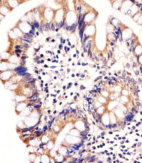
Immunohistochemical of paraffin-embedded H. colon section using CARD6 Antibody. Antibody was diluted at 1:25 dilution. A undiluted biotinylated goat polyvalent antibody was used as the secondary, followed by DAB staining.

CARD6 Antibody western blot of WiDr cell line lysates (35 ug/lane). The CARD6 antibody detected the CARD6 protein (arrow).

Western blot of CARD6 Antibody in MDA-MB231 cell line lysates (35 ug/lane). CARD6 (arrow) was detected using the purified antibody.

Western blot of lysate from WiDr cell line with CARD6 Antibody. Antibody was diluted at 1:1000. A goat anti-rabbit IgG H&L (HRP) at 1:5000 dilution was used as the secondary antibody. Lysate at 20 ug.

Western blot of lysates from A549, HepG2 cell line (from left to right) with CARD6 Antibody. Antibody was diluted at 1:1000 at each lane. A goat anti-rabbit IgG H&L (HRP) at 1:10000 dilution was used as the secondary antibody. Lysates at 20 ug per lane.


