Anti-CDKN2B / p15 INK4b Antibody (aa109-138)
| Name | Anti-CDKN2B / p15 INK4b Antibody (aa109-138) |
|---|---|
| Supplier | LifeSpan Bioscience |
| Catalog | LS-C168624 |
| Prices | $295.00 |
| Sizes | 400 µl |
| Host | Rabbit |
| Clonality | Polyclonal |
| Applications | IHC-P ICC/IF WB FC |
| Species Reactivities | Human |
| Antigen | CDKN2B / p15 INK4b antibody was raised against kLH-conjugated synthetic peptide from C-terminal region of human CDKN2B. |
| Purity/Format | Immunoaffinity purified |
| Blocking Peptide | CDKN2A / p16INK4a Antibody Blocking Peptide |
| Description | Rabbit Polyclonal |
| Gene | CDKN2B |
| Supplier Page | Shop |
Product images
 CDKN2B Antibody immunohistochemistry of formalin-fixed and paraffin-embedded human lung tissue followed by peroxidase-conjugated secondary antibody and DAB staining.
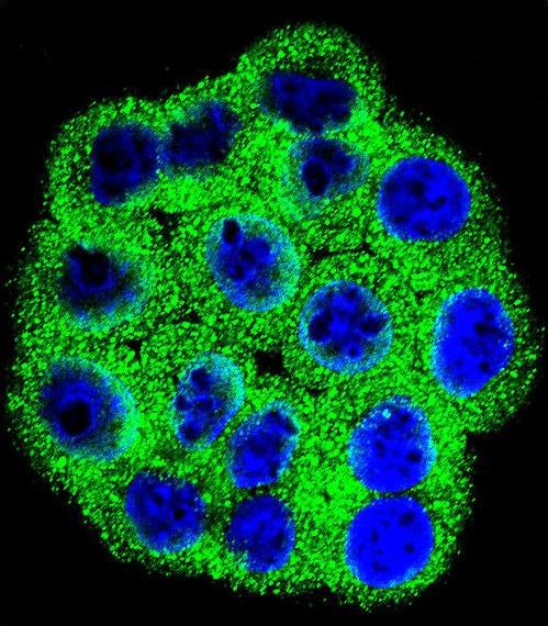 Confocal immunofluorescent of CDKN2B Antibody with HeLa cell followed by Alexa Fluor 488-conjugated goat anti-rabbit lgG (green). DAPI was used to stain the cell nuclear (blue).
 Western blot of CDKN2B (arrow) using rabbit polyclonal CDKN2B Antibody. 293 cell lysates (2 ug/lane) either nontransfected (Lane 1) or transiently transfected (Lane 2) with the CDKN2B gene.
 CDKN2B Antibody flow cytometry of 293 cells (right histogram) compared to a negative control cell (left histogram). FITC-conjugated goat-anti-rabbit secondary antibodies were used for the analysis.