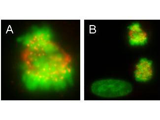
Anti-CENPE Antibody - Immunofluorescence Microscopy. monoclonal anti-CENPE antibody was used to detect CENPE protein, visible as discrete nuclear dots on prometaphase and metaphase cells that relocate to the spindle midzone at anaphase (panel A). Interphase cells show no discrete staining (bottom left, panel B). HeLa cells were fixed in paraformaldehyde and stained using this primary antibody. Alexa Fluor 555 TM conjugated anti-Mouse antibody (red) was used for detection. DNA was stained using bis-benzimide (DAPI) (green). Personal Communication, Tim Yen, Fox Chase Cancer Center, Philadelphia, PA.
