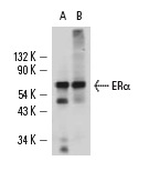
ERα (H226): sc-53493. Western blot analysis of ERα expression in MCF7 nuclear extract (A) and T-47D whole cell lysate (B) under non-reducing conditions.
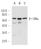
ERα (H226): sc-53493. Western blot analysis of ERα expression in MCF7 nuclear extract (A) and T-47D (B) and ZR-75-1 (C) whole cell lysates.
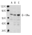
ERα (H226): sc-53493. Western blot analysis of ERα expression in MCF7 nuclear extract (A) and T-47D (B) and ZR-75-1 (C) whole cell lysates.
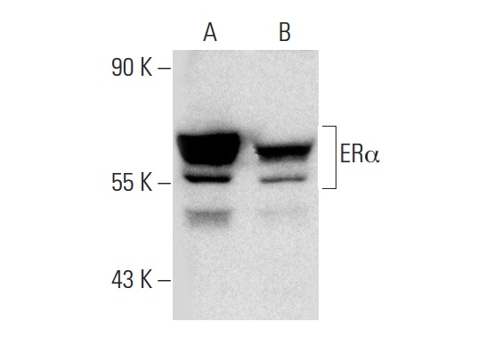
ERα (H226): sc-53493. Western blot analysis of ERα expression in untreated (A) and Radicicol (sc-200620) treated (B) MCF7 whole cell lysates. Note down regulation of ERα expression in lane B.
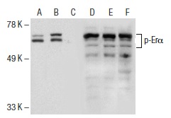
Western blot analysis of ERα phosphorylation in untreated (A,D), estradiol and EGF treated (B,E) and estradiol, EGF and lambda protein phosphatase (sc-200312A) treated (C,F) MCF7 whole cell lysates. Antibodies tested include p-ERα (Ser 118)-R: sc-12915-R (A,B,C) and ERα (H226): sc-53493 (D,E,F).
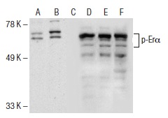
Western blot analysis of ERα phosphorylation in untreated (A,D), estradiol and EGF treated (B,E) and estradiol, EGF and lambda protein phosphatase (sc-200312A) treated (C,F) MCF7 whole cell lysates. Antibodies tested include p-ERα (Ser 118): sc-101675 (A,B,C) and ERα (H226): sc-53493 (D,E,F).
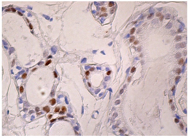
ERα (H226): sc-53493. Immunoperoxidase staining of formalin fixed, paraffin-embedded human breast tissue showing nuclear staining of glandular cells.


