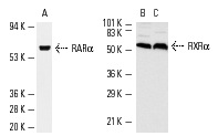
Western blot analysis of RARα (A) and RXRα (B,C) expression in HeLa (A) nuclear extract and NIH/3T3 (B) and KNRK (C) whole cell lysates. Antibodies tested include RARα (C-20): sc-551 (A) and RXRα (D-20): sc-553 (B,C).
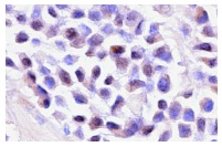
RARα (C-20): sc-551. Immunoperoxidase staining of formalin-fixed, paraffin-embedded human breast tumor showing nuclear and cytoplasmic staining.
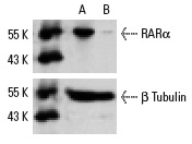
RARα siRNA (h): sc-29465. Western blot analysis of RARα expression in non-transfected control (A) and RARα siRNA transfected (B) HeLa cells. Blot probed with RARα (C-20): sc-551. β Tubulin (D-10): sc-5274 used as specificity and loading control.
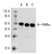
RARα (C-20): sc-551. Western blot analysis of RARα expression in SK-BR-3 (A) and MCF7 (B) nuclear extracts and AML-193 whole cell lysate (C).
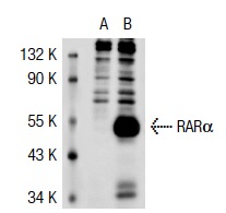
RARα (C-20): sc-551. Western blot analysis of RARα expression in non-transfected: sc-117752 (A) and human RARα transfected: sc-117323 (B) 293T whole cell lysates.
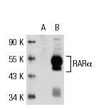
RARα (C-20): sc-551. Western blot analysis of RARα expression in non-transfected: sc-117752 (A) and human RARα transfected: sc-117323 (B) 293T whole cell lysates.
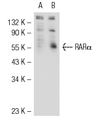
RARα (C-20): sc-551. Western blot analysis of RARα expression in non-transfected: sc-117752 (A) and human RARα transfected: sc-170472 (B) 293T whole cell lysates.
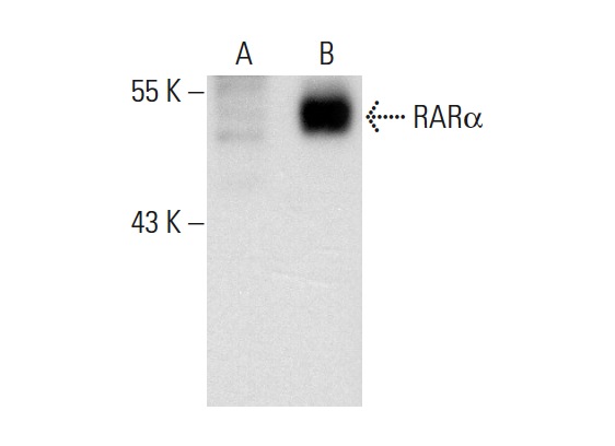
RARα (C-20): sc-551. Western blot analysis of RARα expression in non-transfected: sc-117752 (A) and mouse RARα transfected: sc-125890 (B) 293T whole cell lysates.
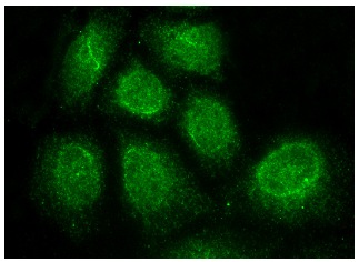
RARα (C-20): sc-551. Immunofluorescence staining of methanol-fixed HeLa cells showing nuclear localization.


