Western blot analysis of extracts from 293T cells, HeLa cells, and human hypothalamus, using GPR50 (D1D6I) Rabbit mAb (upper) and β-Actin (D6A8) Rabbit mAb #8457 (lower).

Immunoprecipitation of GPR50 from 293T cell extracts using Rabbit (DA1E) mAb IgG XP ® Isotype Control #3900 (lane 2) or GPR50 (D1D6I) Rabbit mAb (lane 3). Lane 1 is 10% input. Western blot analysis was performed using GPR50 (D1D6I) Rabbit mAb.

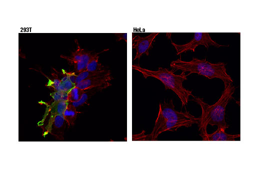
Confocal immunofluorescent analysis of 293T (left, positive), or HeLa (right, negative) cells using GPR50 (D1D6I) Rabbit mAb (green). Actin filaments were labeled with DyLight™ 554 Phalloidin #13054 (red). Blue pseudocolor = DRAQ5 ® #4084 (fluorescent DNA dye).


